A decade-by-decade analysis of how extracellular vesicle science transformed from a niche curiosity into a powerhouse of diagnostics, therapeutics, and precision medicine.
Introduction: A Decade That Redefined Intercellular Communication
A decade ago, extracellular vesicles (EVs) occupied a narrow corner of cell biology — known to a handful of specialists, largely underestimated as mere cellular ‘debris.’ Today, EVs are recognized as fundamental mediators of intercellular communication, disease propagation, and biological signaling that permeates virtually every branch of medicine and life science.
The transformation of EV research between 2015 and 2025 has been nothing short of revolutionary. Driven by advances in nanotechnology, multi-omics, artificial intelligence, and single-particle analysis, the field has moved from descriptive studies of EV existence to mechanistic dissection, clinical biomarker validation, and therapeutic application. Publication output in EV research grew from a few hundred papers per year in 2014 to over 15,000 annually by 2024 — a testament to the field’s explosive momentum.
The International Society for Extracellular Vesicles (ISEV) has been instrumental in shaping this evolution, publishing successive iterations of the Minimal Information for Studies of Extracellular Vesicles (MISEV) guidelines in 2014 and 2018, with a 2023 update anticipated, providing the community with a standardized scientific framework.
This article chronicles the milestones, paradigm shifts, technological breakthroughs, and remaining challenges that have defined EV research’s extraordinary decade of growth.
1. The Foundation Years (2014–2016): From Curiosity to Credibility
The modern EV era arguably began with a series of high-impact publications in 2013–2015 that thrust EVs into the mainstream scientific conversation. Before this period, the EV field suffered from skepticism: were these particles merely cellular waste, or did they carry biologically meaningful cargo?
1.1 The Exosome–Cancer Nexus
A watershed moment arrived in 2015 when Hoshino et al. published a landmark study in Nature demonstrating that tumor-derived exosomes expressing organ-specific integrins could establish pre-metastatic niches — effectively ‘addressing’ future metastatic sites before tumor cell arrival. This single paper redirected the attention of oncology researchers worldwide toward EVs as active architects of cancer biology rather than passive bystanders.
1.2 Glypican-1: The First EV Cancer Biomarker
Also in 2015, Melo et al. reported in Nature that glypican-1 (GPC1)-positive exosomes could distinguish pancreatic cancer patients from healthy controls with remarkable specificity and sensitivity, even at early disease stages. This was one of the first demonstrations that EV surface proteins could serve as non-invasive cancer biomarkers — igniting the liquid biopsy revolution.
1.3 MISEV2014: Setting the Standards
The publication of MISEV2014 by the ISEV established minimum reporting standards for EV studies, addressing the chaos of inconsistent isolation protocols, nomenclature, and characterization methods that had plagued the early field. MISEV2014 mandated rigorous characterization criteria, boosting reproducibility and credibility across research groups globally. The guidelines are accessible at Journal of Extracellular Vesicles.
2. The Technology Surge (2016–2018): Seeing EVs in New Dimensions
The period from 2016 to 2018 was characterized by a technological arms race to characterize EVs with greater resolution, throughput, and specificity. Bulk analysis was giving way to single-particle, multi-parameter platforms that revealed previously invisible EV heterogeneity.
2.1 Nanoparticle Tracking Analysis Goes Mainstream
Nanoparticle tracking analysis (NTA), commercialized through instruments like the NanoSight (Malvern Panalytical), became the de facto standard for EV size distribution and concentration measurement. The ability to track individual particles in real time provided a level of characterization depth previously impossible with bulk methods like dynamic light scattering (DLS).
2.2 Cryo-Electron Microscopy Reveals EV Architecture
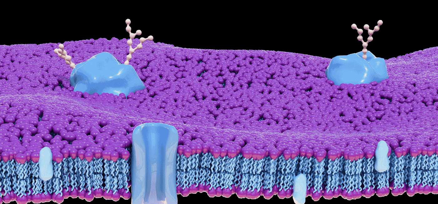
Cryo-EM imaging of EVs provided the first high-resolution structural views of intact EV membranes, surface proteins, and internal cargo organization. These images demolished the notion that EVs were uniform spheres, revealing complex, pleomorphic structures with distinct membrane domains and protein distributions — pointing toward functionally distinct EV subtypes.
2.3 EV Proteomics Matures
Liquid chromatography–tandem mass spectrometry (LC-MS/MS) was increasingly applied to EV proteomics, enabling deep proteomic profiling of hundreds to thousands of EV-associated proteins from complex biological samples. Databases such as Vesiclepedia and EVpedia expanded rapidly, aggregating EV molecular cargo data across cell types, organisms, and disease states to serve as community-wide reference resources.
2.4 MISEV2018: A More Rigorous Framework
Building on MISEV2014, the MISEV2018 guidelines provided updated recommendations for EV isolation, characterization, and functional studies, reflecting the field’s growing sophistication. MISEV2018 introduced more nuanced recommendations about EV subtype classification, urged researchers to use multiple complementary characterization methods, and addressed the problematic overuse of the term ‘exosomes’ for heterogeneous EV populations.
3. The Biomarker Era (2017–2020): EVs Enter the Clinic
With robust characterization tools in hand and a growing catalog of disease-associated EV signatures, researchers turned toward clinical translation. The 2017–2020 period saw the first wave of clinically validated EV biomarker studies across oncology, neurology, and cardiology.
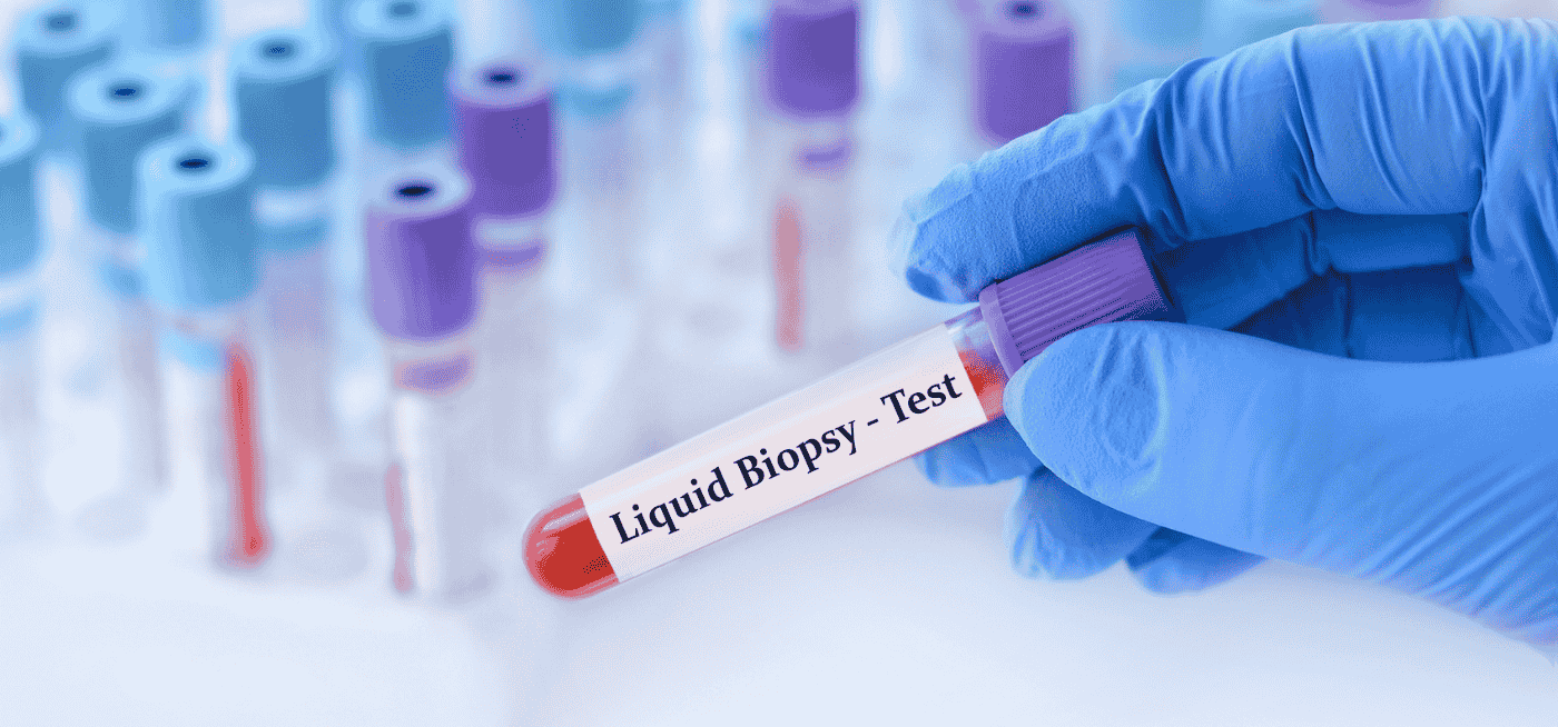
3.1 EV-Based Liquid Biopsy for Cancer
Companies like Exosome Diagnostics launched commercial EV-based liquid biopsy assays, most notably the ExoDx Prostate IntelliScore, which uses EV RNA signatures to refine prostate cancer risk stratification at the time of initial biopsy. This represented one of the first FDA-cleared EV-based diagnostic products, marking a genuine milestone in clinical EV translation.
Beyond prostate cancer, EV liquid biopsy studies were published for lung, breast, colorectal, bladder, and ovarian cancers, demonstrating that plasma or urine-derived EV protein and nucleic acid profiles could detect cancer, classify tumor subtypes, and track treatment response with clinically meaningful performance.
3.2 Neurological Biomarkers: Looking into the Brain Without Surgery
The blood-brain barrier had long blocked easy access to CNS disease biomarkers. A series of studies between 2017 and 2020 showed that neuron-derived exosomes in peripheral blood carry phosphorylated tau, amyloid-β, and neurofilament light chain (NfL) — biomarkers that change years before clinical symptoms of Alzheimer’s and Parkinson’s diseases. For the first time, researchers could interrogate brain pathology through a blood draw.
A notable commercial milestone in this space was the development of NeuroDex, a company pioneering blood-based neurodegenerative disease diagnostics using neuron-derived exosome (NDE) profiling. NeuroDex’s platform isolates NDEs from peripheral blood using neural cell adhesion molecule (NCAM)-based immunocapture, selectively enriching for brain-derived vesicles from the systemic circulation. This approach enables quantification of Alzheimer’s-associated cargo — including phosphorylated tau (p-T181), amyloid-β42, and synaptic proteins — from a standard blood draw, potentially enabling population-scale neurodegenerative disease screening. NeuroDex represents a broader industry trend of translating EV neurobiology research into accessible, minimally invasive clinical tools that sidestep the cost and invasiveness of cerebrospinal fluid collection or PET imaging for CNS disease monitoring.
3.3 Urinary EV Profiling for Kidney and Bladder Disease
Urine is an ideal, non-invasive EV source for urological and renal disease monitoring. Studies during this period validated urinary EV signatures for early detection of acute kidney injury, diabetic nephropathy, bladder cancer, and urinary tract infection — offering the prospect of completely non-invasive kidney disease monitoring at scale.
3.4 EV-TRACK: Transparent Reporting Standards
The EV-TRACK platform was launched in 2017 as a community-driven knowledgebase to promote transparent reporting of EV experiments. Researchers could deposit experimental metadata alongside publications, enabling readers to assess methodological rigor and enabling meta-analyses of EV biomarker studies. EV-TRACK became a model for data transparency in biomedical research.
4. The COVID-19 Interlude (2020–2021): EVs in a Global Pandemic
The COVID-19 pandemic created an unexpected but enormously informative chapter in EV research. SARS-CoV-2 infection profoundly dysregulated EV biogenesis, and EV profiling emerged as a powerful tool for understanding COVID-19 pathogenesis, severity prediction, and long-COVID biology.
4.1 SARS-CoV-2 Hijacks EV Biogenesis
Multiple research groups demonstrated that SARS-CoV-2 exploits host EV machinery to package viral proteins — including spike, envelope, and nucleocapsid — into EV-like particles. These virus-mimicking particles could transfer viral components to uninfected cells, potentially amplifying infection, immune evasion, and systemic inflammation.
4.2 EV Profiling Illuminates COVID-19 Severity
Proteomic and lipidomic profiling of plasma EVs from COVID-19 patients revealed dramatic upregulation of coagulation factors, complement proteins, pro-inflammatory cytokines, and endothelial activation markers — molecular fingerprints that correlated with disease severity, ICU admission, and mortality. EV profiling thus provided mechanistic insights into the cytokine storm and endotheliopathy underlying severe COVID-19.
4.3 Therapeutic EVs for Lung Injury
The respiratory devastation of COVID-19 accelerated interest in mesenchymal stem cell (MSC)-derived EV therapy for acute lung injury. Multiple clinical trials were initiated using MSC-EVs as anti-inflammatory agents in severe COVID-19 pneumonia, providing real-world safety and preliminary efficacy data that will benefit EV therapeutics broadly beyond the pandemic context.
5. The Multi-Omics Revolution (2020–2023): Decoding EV Complexity
As single-omics EV studies proliferated, it became clear that no single molecular layer — proteome, transcriptome, or lipidome alone — could fully capture EV biology. The 2020–2023 period was defined by the rise of integrated multi-omics EV profiling and the deployment of artificial intelligence to synthesize this complexity.
5.1 Single-EV Multi-Parameter Analysis
Groundbreaking single-EV analysis platforms, including the ExoView (NanoView Biosciences) and nano-flow cytometry systems, enabled simultaneous profiling of multiple surface proteins on individual EVs. These technologies revealed that EV populations once assumed to be homogeneous were in fact composed of functionally distinct subpopulations with non-overlapping protein signatures — a discovery with profound implications for disease biomarker specificity and therapeutic targeting.
5.2 EV Transcriptomics: Beyond miRNA
Early EV RNA studies focused almost exclusively on miRNAs. The 2020–2023 period saw an expansion into EV long non-coding RNAs (lncRNAs), circular RNAs (circRNAs), and even full-length mRNAs. CircRNA-enriched EVs emerged as particularly stable and abundant cargo with roles in cancer progression and immune regulation, opening an entirely new dimension of EV biology.
5.3 Artificial Intelligence Decodes EV Signatures
Machine learning and deep learning algorithms were applied to large EV multi-omics datasets, enabling disease classification, biomarker panel selection, and pathway analysis at a scale and depth impossible with conventional statistics. AI-driven EV proteomic classifiers achieved high accuracy in distinguishing cancer types, treatment-resistant tumors, and early-stage lesions — accelerating the path from discovery to clinical assay development.
5.4 Spatial Biology Meets EV Research
Spatial transcriptomics and proteomics technologies, originally developed for tissue biology, began to be coupled with EV research — mapping where EVs are produced, released, and taken up within intact tissue and tumor architectures. This spatial dimension added a crucial layer of context to EV molecular profiles, revealing how tissue geography shapes EV signaling networks in disease.
6. The Therapeutic Decade (2018–2025): EVs as Medicines
Arguably the most consequential development of the last decade has been the emergence of EVs as a new class of therapeutic agents — both as natural anti-inflammatory biologics and as engineered drug delivery vehicles.
6.1 MSC-Derived EVs: Regenerative Medicine Without the Cells
Mesenchymal stem cell (MSC)-derived EVs recapitulate many of the regenerative, anti-inflammatory, and immunomodulatory properties of their parent cells — without the risks of live-cell therapy including immunogenicity, oncogenic potential, and vascular occlusion. Between 2018 and 2025, MSC-EV therapies entered clinical trials for graft-versus-host disease, acute respiratory distress syndrome, myocardial infarction, and neurodegenerative disease, with several Phase II trials reporting encouraging safety and efficacy signals.
6.2 Engineered EVs: Precision Drug Delivery
Engineered EVs — modified to display targeting ligands, receptor-binding peptides, or therapeutic payloads — advanced from proof-of-concept animal studies to early human trials. Loading strategies including electroporation, sonication, and co-extrusion were optimized to package siRNA, CRISPR-Cas9 complexes, chemotherapy, and mRNA into EVs with high efficiency and minimal cargo degradation. A landmark 2021 study in Nature Biomedical Engineering demonstrated targeted EV delivery of siRNA to KRAS-mutant pancreatic cancer in vivo — a historically undruggable oncogene.
6.3 EV Vaccines: A New Immunization Paradigm
Bacterial outer membrane vesicle (OMV)-based vaccines have been clinically approved for meningococcal disease for over a decade. The last five years saw intense development of eukaryotic EV vaccines — particularly tumor antigen-loaded exosome vaccines for cancer immunotherapy. Phase I trials in melanoma and non-small cell lung cancer demonstrated safety and preliminary evidence of antigen-specific immune responses, with Phase II trials now underway.
6.4 Plant-Derived EVs: An Unexpected Frontier
An unexpected discovery of the decade was that plants secrete nanoparticle-like EVs that, when consumed or administered systemically, exert anti-inflammatory and gut-protective effects in mammalian systems. Plant-derived EVs from ginger, grapes, and grapefruit were shown to reduce colitis, protect against alcohol-induced liver injury, and modulate the gut microbiome — opening a pharmacognosy-inspired dimension of EV therapeutics with natural, scalable production advantages.
7. Regulatory and Standardization Milestones
Translating EV science into clinical products required not just biological breakthroughs but also regulatory clarity and manufacturing standardization — areas that matured considerably over the decade.
7.1 FDA Engagement with EV Therapeutics
The U.S. Food and Drug Administration (FDA) issued guidance documents addressing the characterization, potency assays, and manufacturing standards for EV-based cell and gene therapy products. The FDA’s Center for Biologics Evaluation and Research (CBER) engaged with EV therapeutic developers through the Regenerative Medicine Advanced Therapy (RMAT) designation pathway, accelerating clinical development timelines for promising EV drug candidates.
7.2 Good Manufacturing Practice (GMP) for EVs
Producing therapeutic-grade EVs at clinical scale demanded the development of GMP-compliant manufacturing workflows — scalable bioreactor-based EV production, standardized quality control assays, and long-term stability data. Major academic medical centers and contract development and manufacturing organizations (CDMOs) invested heavily in EV GMP capabilities, with several establishing dedicated EV manufacturing suites by 2023–2024.
7.3 EV Reference Materials and Calibrants
The National Institute of Standards and Technology (NIST) and the ISEV collaborated to develop reference EV materials — standardized nanoparticle preparations with known size, concentration, and protein profiles — enabling inter-laboratory comparability for the first time. These reference standards are foundational for regulatory submissions and diagnostic assay validation.
8. Key Paradigm Shifts of the Decade
Beyond individual discoveries, the last decade produced several fundamental paradigm shifts that have reshaped how the scientific community conceptualizes EVs:
- From waste to signal: EVs transitioned from being considered cellular debris to being understood as highly regulated, information-rich signaling particles that orchestrate tissue homeostasis and disease.
- From homogeneity to heterogeneity: The assumption that EV populations are uniform gave way to appreciation of functionally distinct EV subpopulations with unique molecular identities and biological activities.
- From bulk to single-particle: Analytical approaches shifted from ensemble measurements averaging across millions of EVs to single-particle resolution, revealing EV diversity invisible to bulk methods.
- From exosomes to EVs: The term ‘exosome’ was increasingly recognized as imprecise for heterogeneous EV preparations. The field converged on ‘extracellular vesicles’ as the appropriate umbrella term for rigorously characterized preparations.
- From passive cargo to active programming: EVs are now understood to actively reprogram recipient cell gene expression, metabolism, and signaling — not merely deliver molecular messages passively.
- From research tool to clinical product: EVs evolved from research curiosities to FDA-engaged therapeutic and diagnostic products entering clinical trials worldwide.
9. Remaining Challenges as the Decade Closes
Despite extraordinary progress, the EV field enters its next decade with significant challenges that remain partially or fully unresolved.
9.1 Heterogeneity and Subpopulation Isolation
Even with single-EV analysis tools, selectively isolating pure, functional EV subpopulations from complex biological fluids remains technically demanding and incompletely standardized. Without reliable subpopulation isolation, mechanistic studies and biomarker discovery remain confounded by mixed EV signals.
9.2 In Vivo EV Tracking
Understanding EV biodistribution, targeting efficiency, and fate in living organisms remains challenging. Current in vivo imaging approaches — including fluorescent labeling and bioluminescence — perturb EV properties and lack single-EV resolution in deep tissues. Next-generation EV tracking tools are urgently needed.
9.3 Scalable GMP Manufacturing
Producing the quantities of therapeutic-grade EVs required for clinical trials and eventual commercialization remains a bottleneck. Bioreactor-based production, hollow-fiber filtration, and alternative EV sources are being actively optimized, but scalability, batch-to-batch consistency, and cost-effectiveness remain obstacles.
9.4 Long-Term Clinical Evidence
Most EV biomarker studies remain cross-sectional and exploratory. Longitudinal clinical validation cohorts with thousands of patients, multi-site standardization, and clinical outcome correlation are needed before EV diagnostics achieve the evidence level required for widespread clinical adoption.
10. The Road Ahead: EV Research in the Next Decade
The next decade of EV research will likely be defined by convergence — between basic biology and clinical medicine, between EV profiling and other liquid biopsy modalities, and between EV therapeutics and precision medicine.
Multi-modal liquid biopsy panels combining EV protein signatures, EV-derived nucleic acids, cell-free DNA (cfDNA), and circulating tumor cells (CTCs) are likely to outperform any single analyte in clinical sensitivity and specificity. EV profiling will also integrate with single-cell sequencing data to map EV-mediated intercellular communication networks at tissue and organismal scale — an ambition now tractable with technologies like 10x Genomics Visium and related spatial multi-omics platforms.
On the therapeutic front, engineered EV drugs targeting previously intractable targets — mutant RAS, tau aggregates, CNS tumors — are expected to advance through Phase II and III clinical trials. The regulatory landscape will mature, with established GMP standards, potency assays, and clinical pharmacology frameworks enabling faster and more predictable approval pathways.
Ultimately, the next decade will determine whether EV research’s extraordinary promise translates into transformative clinical impact — earlier cancer detection, disease-modifying neurological therapies, and regenerative medicines that fundamentally change patient outcomes.
Conclusion: A Decade That Changed Everything
The evolution of EV research over the last decade represents one of the most remarkable transformations in modern biomedical science. From contested biological curiosities to validated disease biomarkers, from cell culture experiments to Phase II clinical trials, and from rudimentary ultracentrifugation to AI-powered single-particle multi-omics — EVs have arrived at the center of 21st-century medicine.
The scientific community’s investment in standardization, technology development, and clinical translation has created a field that is simultaneously rigorous and dynamic. The next decade will determine whether this momentum delivers on its extraordinary promise — and all indicators suggest that it will.
References
[1] Théry C, et al. (2018). MISEV2018 guidelines for EV studies. Journal of Extracellular Vesicles.
[3] Hoshino A, et al. (2015). Tumour exosome integrins determine organotropic metastasis. Nature.
[6] Pegtel DM, Gould SJ. (2019). Exosomes. Annual Review of Biochemistry.
[8] Vader P, et al. (2016). Extracellular vesicles for drug delivery. Advanced Drug Delivery Reviews.
[12] Vesiclepedia — Comprehensive EV Molecular Cargo Database.
[13] Exosome Diagnostics — ExoDx Prostate IntelliScore.
[15] FDA CBER — Center for Biologics Evaluation and Research.