
A translational biomarker team building a plasma panel for a neurodegeneration trial usually starts with the same three candidates: GFAP for astrocytic injury, NfL for axonal damage, and increasingly, TDP-43 for disease-specific proteinopathy. Each has a defensible rationale. Put them in the same whole-plasma panel, though, and the team inherits three different specificity problems at once — and for TDP-43 in particular, the published evidence doesn’t yet agree with itself. This piece looks at what changes, marker by marker, when the same three analytes are measured from a neuron-derived extracellular vesicle (NDEV) fraction instead of whole plasma.
Key Takeaways
- GFAP and NfL are genuinely validated blood-based markers of astrocytic and axonal injury, but neither is specific to a disease mechanism or to CNS origin.
- Whole-plasma TDP-43 has produced contradictory findings across cohorts — null in a large population-based study, positive in a disease-enriched autopsy cohort — a specificity problem, not a data-quality one [1,2].
- A 2025 perspective on plasma GFAP explicitly raises blood-brain-barrier origin uncertainty and proposes astrocyte-derived exosome analysis as one path toward resolving it [3].
- Multiplexing markers from an NDEV-enriched fraction gives each analyte a shared CNS-origin filter, rather than asking three different proteins to each individually overcome peripheral background.
- Rigor in isolation and characterization method — per MISEV2023 — is the precondition for any of this being usable in a translational program, not an afterthought [4].
GFAP and NfL Have Earned Their Place — With a Real Asterisk
Neurofilament light chain is the most clinically validated blood-based marker of neuroaxonal injury in ALS and several other neurodegenerative conditions, and a study correlating NfL, TDP-43, and total tau across CSF and plasma in 75 ALS patients found NfL tracked with disease progression rate in multivariate analysis [5]. GFAP has a comparably strong track record for astrocytic reactivity, with elevated plasma levels associated with amyloid burden, tau pathology, and neurodegeneration in Alzheimer’s disease cohorts.
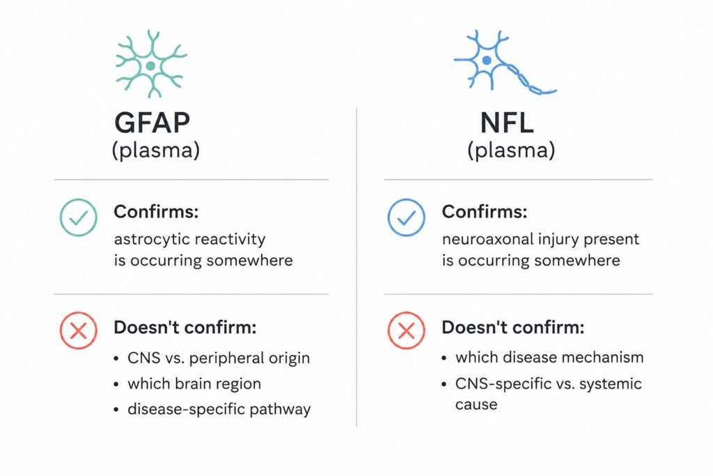
Both markers deserve the confidence the field has placed in them. Neither one, though, tells a translational team what they often actually need to know: which disease-specific pathology is driving the signal, and whether it originated in the brain at all. NfL rises with axonal injury from essentially any cause. And a 2025 perspective piece on plasma GFAP is unusually direct about the second problem — the authors note that the tissue origin of blood GFAP remains genuinely unclear, and that it isn’t settled whether it derives from CSF efflux or specifically from reactive astrocytes, particularly in neurodegeneration where blood-brain barrier integrity is often preserved [3]. Their proposed path forward is notable: analyzing astrocyte-derived exosomes as a way to attribute the signal to its cellular source, rather than relying on whole-fluid measurement alone [3].
Figure 1. The specificity gap: what GFAP and NfL confirm vs. what they can’t
TDP-43: A Marker Whose Own Evidence Base Disagrees With Itself
TDP-43 is the more mechanistically specific marker of the three — it’s a hallmark pathology in ALS, FTD, and limbic-predominant age-related TDP-43 encephalopathy (LATE) — but the whole-plasma evidence for measuring it is genuinely inconsistent in a way that’s worth stating plainly rather than glossing over.
In a population-based cohort of 1,058 participants in the Cardiovascular Health Study, plasma TDP-43 was not associated with cognitive decline, incident dementia, plasma AD biomarkers, or brain MRI volumes over a mean 5.5-year follow-up [1]. In a separate study using an ultrasensitive immunoassay on samples from autopsy-confirmed cases, plasma TDP-43 and phospho-TDP-43 were significantly elevated in advanced LATE neuropathologic change, with an ROC area under the curve approaching 0.8 in the subgroup with comorbid Alzheimer’s pathology [2]. A systematic review and meta-analysis of CSF TDP-43 across seven pooled ALS studies similarly concluded the marker shows promise but that further studies are needed before firm conclusions can be drawn [6].
These aren’t competing claims about the same result — they’re consistent with a single explanation. TDP-43 is expressed outside the CNS, and a whole-plasma sample captures TDP-43 shed from any tissue. How much that non-neuronal background dilutes the signal will vary by cohort composition, disease stage, and comorbidity burden, which is a reasonable account of why a population-based cohort and a disease-enriched autopsy cohort reached different conclusions using the same analyte [1,2].
What a Shared CNS-Origin Filter Actually Changes
A 2024 review examining brain-derived extracellular vesicles within the Research Domain Criteria (RDoC) framework makes the case for BDEVs as non-invasive mechanistic biomarkers precisely because their cargo — protein and RNA — reflects the biology of the cell that released them [7]. That review is candid that isolation method, vesicle characterization, and cargo-readout standardization all still need further work across the field before BDEV-based biomarkers can support high-stakes drug development decisions [7].
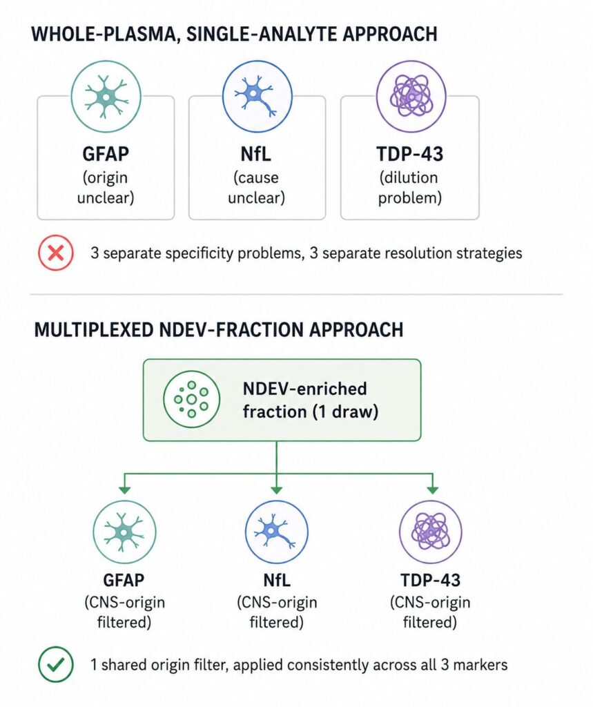
The practical implication for a three-marker panel is straightforward: instead of asking GFAP to somehow indicate astrocyte-of-origin on its own, asking NfL to somehow indicate CNS-of-origin on its own, and asking TDP-43 to somehow overcome dilution from every other tissue that expresses it, all three markers get measured from a fraction that’s already been filtered to neuron-derived cargo before quantification. The GFAP perspective piece essentially proposed this solution for one marker in isolation — astrocyte-derived exosome analysis as a way to resolve GFAP’s origin question [3]. Multiplexing across an NDEV-enriched fraction extends the same logic to all three analytes from a single plasma draw, rather than requiring a separate resolution strategy per marker.
Figure 2. Single-analyte specificity problem vs. multiplexed NDEV-fraction approach
Comparing the Three Markers Head-to-Head
| Marker | Established strength | Documented limitation | What NDEV multiplexing adds |
|---|---|---|---|
| GFAP | Strong association with astrocytic reactivity across AD cohorts | Blood-brain-barrier origin uncertain even when BBB integrity is preserved [3] | Attributes signal to CNS cell-of-origin before quantification |
| NfL | Best-validated blood marker of neuroaxonal injury in ALS [5] | Reflects general axonal damage, not disease-specific mechanism | Same origin filter, applied alongside disease-specific markers |
| TDP-43 | Mechanistically specific to ALS/FTD/LATE pathology | Whole-plasma findings contradict across cohorts [1,2] | Reduces non-neuronal dilution before the assay runs |
Table is illustrative, synthesized from the cited primary literature rather than a single head-to-head study.
Rigor Comes First, Not After
None of this is a substitute for standardization discipline. MISEV2023 — the current consensus guideline from the International Society for Extracellular Vesicles — lays out the isolation, characterization, and reporting standards that any EV-based biomarker measurement needs to meet, updating and extending the field’s earlier 2018 guidance [4]. A multiplex NDEV panel that skips defined vesicle markers, documented isolation method, and reproducibility metrics doesn’t solve the specificity problems described above — it just moves them somewhere less visible. The value of an NDEV-enriched fraction depends entirely on the isolation step being done to that standard.
The field’s evidence base on all three of these markers is still developing, and the GFAP and TDP-43 literature reviewed here is a useful reminder that whole-plasma measurement alone hasn’t settled several open questions. What multiplexing across a properly characterized NDEV fraction changes is the starting point — giving GFAP, NfL, and TDP-43 a shared, CNS-attributable basis before any of them are asked to answer a disease-specific question. The LuminEV Research Kit is built around that multiplex, one-sample approach, running these markers together from a single plasma draw within a defined, standardized workflow.
References
[1] Fohner AE, Sitlani CM, Jayadev S, Bis JC, Trittschuh EH, Lopez OL, Tracy RP, Psaty BM, Longstreth WT Jr, Seshadri S. Plasma TAR DNA-binding protein 43 (TDP-43) levels in a population-based cohort of older adults: The Cardiovascular Health Study. J Alzheimers Dis. 2025;105(4):1275-1281. https://doi.org/10.1177/13872877251334820
[2] Wang J, Schneider JA, Bennett DA, Seyfried NT, Young-Pearse TL, Yang HS. Plasma TDP-43 is a potential biomarker for advanced limbic-predominant age-related TDP-43 encephalopathy neuropathologic change. Mol Neurodegener. 2025;20:119. https://doi.org/10.1186/s13024-025-00910-4
[3] Youn W, Yun M, Lee CJ, Schöll M. Cautions on utilizing plasma GFAP level as a biomarker for reactive astrocytes in neurodegenerative diseases. Mol Neurodegener. 2025;20:54. https://doi.org/10.1186/s13024-025-00846-9
[4] Welsh JA, Goberdhan DCI, O’Driscoll L, et al. Minimal information for studies of extracellular vesicles (MISEV2023): From basic to advanced approaches. J Extracell Vesicles. 2024;13(2):e12404. https://doi.org/10.1002/jev2.12404
[5] Kojima Y, Kasai T, Noto Y, Ohmichi T, Tatebe H, Kitaoji T, Tsuji Y, Kitani-Morii F, Shinomoto M, Allsop D, Teramukai S, Mizuno T, Tokuda T, Le W. Amyotrophic lateral sclerosis: Correlations between fluid biomarkers of NfL, TDP-43, and tau, and clinical characteristics. PLoS One. 2021;16(11):e0260323. https://doi.org/10.1371/journal.pone.0260323
[6] Gambino CM, Ciaccio AM, Lo Sasso B, Giglio RV, Vidali M, Agnello L, Ciaccio M. The role of TAR DNA binding protein 43 (TDP-43) as a candidate biomarker of amyotrophic lateral sclerosis: a systematic review and meta-analysis. Diagnostics (Basel). 2023;13(3):416. https://doi.org/10.3390/diagnostics13030416
[7] Magaraggia I, Krauskopf J, Ramaekers JG, You Y, de Nijs L, Briedé JJ, Schreiber R. Harnessing brain-derived extracellular vesicles to support RDoC-based drug development. Neuroscience Applied. 2024;4:105406. https://doi.org/10.1016/j.nsa.2024.105406