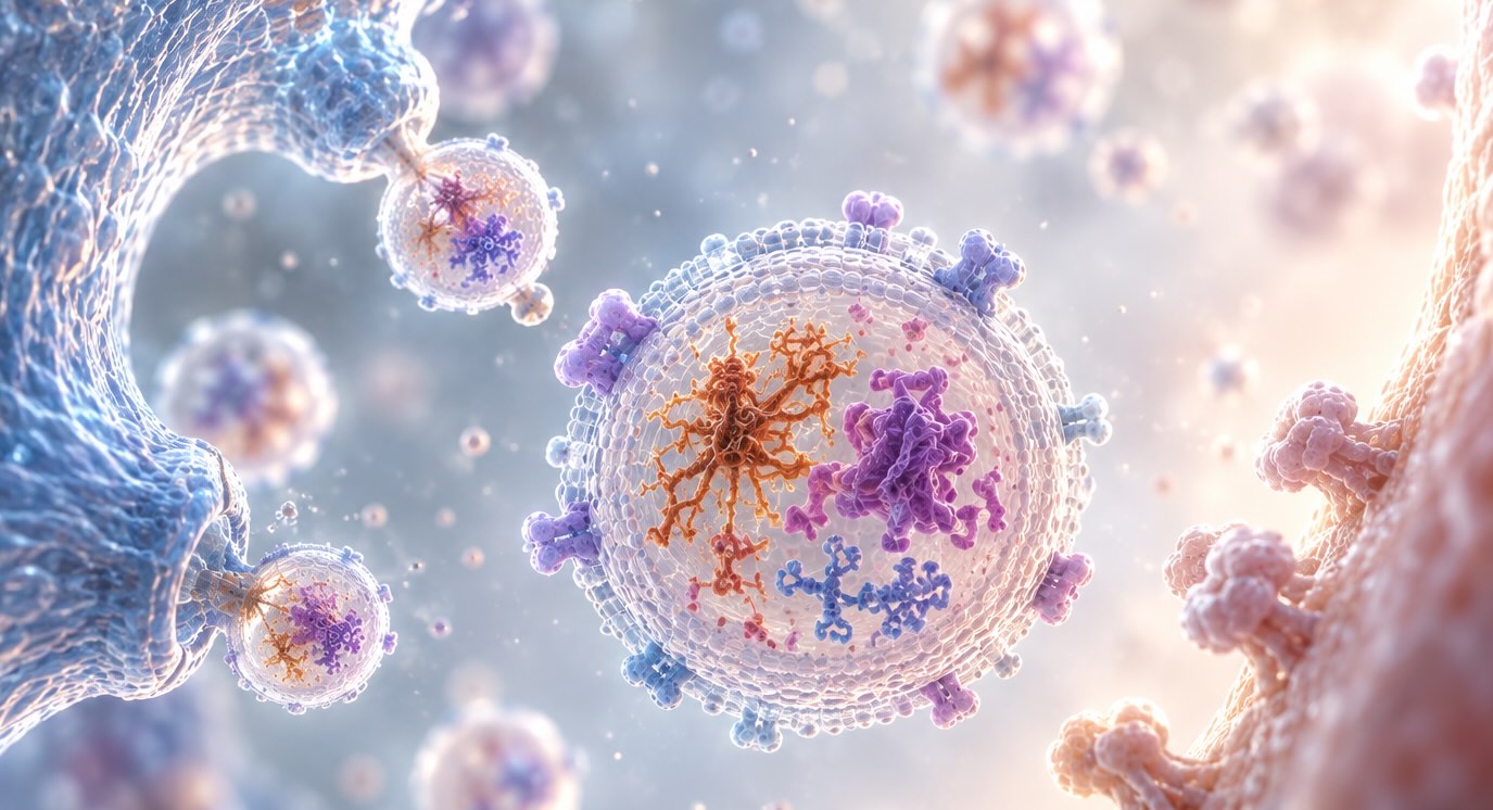


A researcher with 200 µL of plasma and five candidate markers has already lost the study before the first plate is read. Split five ways across single-analyte assays, that volume barely covers duplicates — and that’s before accounting for dead volume, repeat runs, and the QC failures that come with any single plate. The problem isn’t the biology. It’s that the assay format was built to answer one question at a time, in a field where the disease itself never asks just one.
Neurodegeneration doesn’t proceed marker by marker. Tau, TDP-43, α-synuclein, and glial fibrillary acidic protein (GFAP) co-occur, correlate, and interact across Alzheimer’s, Parkinson’s, ALS, and frontotemporal lobar degeneration. A plasma sample run through a single-analyte pipeline gives back one thread of that picture — and consumes the sample needed to pull the rest.
Key Takeaways
- Single-analyte assays scale sample consumption and hands-on time linearly with every marker added, a math problem that becomes untenable once a panel exceeds two or three targets.
- Sequential, plate-by-plate testing of individual markers introduces batch-to-batch and lot-to-lot variability that a co-run panel avoids by design.
- Extracellular vesicle (EV) surface and cargo markers relevant to neurodegeneration rarely act alone — plasma NfL, GFAP, and total tau are correlated in Parkinson’s cohorts, and synaptic panels distinguish TDP-43 from tau pathology only when multiple markers are read together.
- Multiplexed bead-based platforms have been validated against single-analyte formats for reproducibility, linearity, and recovery — the technology is not new, but its adoption in EV biomarker workflows lags behind its adoption in cytokine profiling.
The Sample Volume Math No One Budgets For
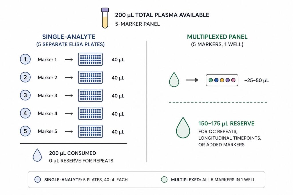
Plasma is not a renewable resource within a study. Once a low-volume draw is centrifuged, aliquoted, and frozen, the researcher is working against a fixed ceiling — and every single-analyte ELISA plate claims a fixed share of it, plus dead volume for pipetting error, plus enough for repeats when a plate fails QC.
This isn’t a hypothetical constraint. Methods developed specifically to conserve EV-associated material for multi-parametric analysis report protocols built around as little as 1–4 mL of plasma total — for DNA, RNA, and protein extraction combined [4]. That’s the budget an EV biomarker study has to work with when it wants more than one readout per sample. A workflow that spends that budget one analyte at a time, on separate plates, forecloses the rest of the panel before the science even starts.
The dynamic range problem compounds this. Multiplex bead-based platforms have demonstrated linearity across nearly the full concentration range needed for low- and high-abundance analytes within a single well, using a fraction of the sample volume traditional ELISA requires per analyte [2]. A study designed around single-analyte testing isn’t just slower — it’s structurally incapable of testing as many markers per patient as a multiplexed design, at any budget.
Figure 1. Sample Volume Consumption: Single-Analyte vs. Multiplexed Panel
Batch Effects Are a Design Flaw, Not a Fluke
Running five markers across five separate plates means five separate calibration curves, five sets of reagent lots, five incubation cycles — each one a fresh opportunity for plate-to-plate drift. When those markers are meant to be interpreted together, as a ratio or a composite score, that drift becomes signal contamination. The researcher isn’t just measuring biology anymore; they’re measuring which day each plate was run.
This is a known and well-characterized problem in immunoassay validation. Comparative work across commercial multiplex platforms has shown that co-running analytes in a shared assay format avoids the added imprecision that comes from testing the same samples across independent plate runs, and multiplex formats have been shown to meet or exceed the accuracy and reproducibility benchmarks required for biomarker validation studies at scale [2,3]. The rigor bar single-analyte ELISA is held to — precise calibration curves, controlled recoveries, validated linearity — has already been cleared by multiplex bead-based formats built for exactly this kind of parallel analysis [3].
For EV biomarker panels specifically, this matters more than it does for a single soluble protein. Isolation-free multiplex assays built for EV surface protein detection have been directly validated against simplex (single-analyte) runs on the same platform, comparing linearity, spike-in recovery, and inter- and intra-assay precision between the two formats [7]. The finding wasn’t that multiplexing sacrifices precision to gain throughput — it was that a well-designed multiplex panel holds precision steady while removing the batch-to-batch variability that sequential single-analyte testing introduces by design.
The Biology Was Never Single-Marker to Begin With
Even setting sample volume and batch effects aside, there’s a more basic argument against single-analyte testing: the underlying pathology doesn’t isolate itself to one protein.
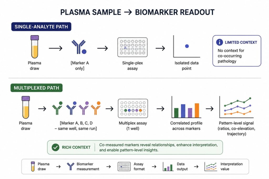
In a cross-sectional Parkinson’s cohort assessed with a four-marker plasma panel, NfL levels correlated significantly with both GFAP and α-synuclein — relationships that would be invisible to a study measuring NfL alone [5]. None of the four individual markers reliably distinguished PD patients from controls on their own; what tracked with motor severity and disease stage was the pattern across markers assessed in the same samples, using the same assay run, at the same time [5].
The same logic holds in frontotemporal lobar degeneration, where a multimarker CSF synaptic protein panel was needed to distinguish FTLD-tau from FTLD-TDP pathology — a distinction that improved diagnostic performance only when synaptic markers were read as a panel rather than individually, and that also tracked with cognitive performance in ways single markers alone could not achieve [6]. Neurodegenerative co-pathology is the norm, not the exception; a testing format that can only afford to look at one marker per plasma draw is, by construction, blind to it.
Figure 2. Where Single-Analyte Testing Loses the Signal
What a Multiplexed Workflow Actually Buys a Study
None of this is an argument that multiplexing is a novel idea — bead-based multiplex immunoassays have been part of the biomarker discovery toolkit since well before EV research adopted them widely [2]. What’s changed is their extension to EV-specific surface and cargo markers, where isolation-free, multiplexed detection formats now allow researchers to profile several EV-associated proteins from unprocessed plasma in a single run, without the sequential, plate-by-plate testing that single-analyte ELISA requires [7].
For a translational biomarker program working across Alzheimer’s, Parkinson’s, ALS, and FTD, that shift changes what a study can ask. Instead of designing a discovery cohort around the one marker with the most literature support and hoping it holds up, a multiplexed EV panel lets a study test tau, TDP-43, α-synuclein, and GFAP-associated signal together, from the same limited plasma draw, in the same assay run — preserving both sample and statistical power for the co-occurring biology that single-marker testing was never built to see.
LuminEV was designed around this constraint directly: multiplexed, isolation-free profiling of EV surface markers from a single plasma aliquot, built to preserve sample volume across a panel rather than consume it one analyte at a time.
Table 1. Single-Analyte vs. Multiplexed EV Panel — Side-by-Side
| Factor | Single-Analyte ELISA | Multiplexed Bead-Based Panel |
|---|---|---|
| Plasma volume per marker | Fixed per plate; scales linearly with panel size | Shared across markers in one well |
| Plate-to-plate batch variability | Present — separate curves, runs, lots per marker | Minimized — markers read in the same run |
| Cross-marker correlation data | Not directly comparable across separate runs | Native to the assay — markers co-measured |
| Throughput per sample | One result per plate | Multiple results per well |
| Validated against single-analyte performance | N/A (is the reference format) | Yes — linearity, recovery, precision benchmarked directly [7] |
Where This Leaves Study Design
The choice between single-analyte and multiplexed testing isn’t a matter of preference once a panel exceeds two markers and the biology under study is known to involve more than one pathological process. Sample volume is finite, batch effects are cumulative, and the co-occurring markers most relevant to Alzheimer’s, Parkinson’s, ALS, and FTD research don’t resolve into single-variable answers. The question worth asking before a study is designed isn’t which single marker to prioritize — it’s whether the assay format chosen can afford to measure them all from what the patient actually gave.
The next step for the field isn’t more single-marker validation studies; it’s building reference ranges and reproducibility data for multiplexed EV panels at the scale that CSF proteomics and cytokine profiling already have. That standardization work is underway, but it’s earlier than the underlying assay technology deserves.
References
[1] Théry C, Witwer KW, Aikawa E, et al. Minimal information for studies of extracellular vesicles 2018 (MISEV2018): a position statement of the International Society for Extracellular Vesicles and update of the MISEV2014 guidelines. J Extracell Vesicles. 2018;7(1):1535750. https://doi.org/10.1080/20013078.2018.1535750
[2] Fu Q, Zhu J, Van Eyk JE. Comparison of Multiplex Immunoassay Platforms. Clin Chem. 2010;56(2):314–318. https://doi.org/10.1373/clinchem.2009.135087
[3] Bastarache JA, Koyama T, Wickersham NE, Ware LB. Accuracy and reproducibility of a multiplex immunoassay platform: A validation study. J Immunol Methods. 2011;367(1–2):33–39. https://doi.org/10.1016/j.jim.2011.01.005
[4] Roy JW, Taylor CA, Beauregard AP, Dhadi SR, Ayre DC, Fry S, Chacko S, Wajnberg G, Joy AP, Mai-Thi NN, Crapoulet N, Barnett DA, Ghosh A, Lewis SM, Ouellette RJ. A multiparametric extraction method for Vn96-isolated plasma extracellular vesicles and cell-free DNA that enables multi-omic profiling. Sci Rep. 2021;11(1):8085. https://doi.org/10.1038/s41598-021-87526-y
[5] Youssef P, Hughes L, Kim WS, Halliday GM, Lewis SJG, Cooper A, Dzamko N. Evaluation of plasma levels of NFL, GFAP, UCHL1 and tau as Parkinson’s disease biomarkers using multiplexed single molecule counting. Sci Rep. 2023;13:5217. https://doi.org/10.1038/s41598-023-32480-0
[6] Cervantes González A, Irwin DJ, Alcolea D, McMillan CT, Chen-Plotkin A, Wolk D, Sirisi S, Dols-Icardo O, Querol-Vilaseca M, Illán-Gala I, Santos-Santos MA, Fortea J, Lee EB, Trojanowski JQ, Grossman M, Lleó A, Belbin O. Multimarker synaptic protein cerebrospinal fluid panels reflect TDP-43 pathology and cognitive performance in a pathological cohort of frontotemporal lobar degeneration. Mol Neurodegener. 2022;17:29. https://doi.org/10.1186/s13024-022-00534-y
[7] Tordoff E, Allen J, Elgart K, Elsherbini A, Kalia V, Wu H, Eren E, Kapogiannis D, Gololobova O, Witwer K, Volpert O, Eitan E. A novel multiplexed immunoassay for surface-exposed proteins in plasma extracellular vesicles. J Extracell Vesicles. 2024;13(11):e70007. https://doi.org/10.1002/jev2.70007