
A comprehensive guide to extracellular vesicle profiling, its role in disease research, and its transformative potential in diagnostics, therapeutics, and precision medicine.
Introduction: The Rise of Extracellular Vesicle Research
Extracellular vesicles (EVs) have emerged as one of the most exciting frontiers in modern biomedical research. These nanoscale, membrane-enclosed particles are secreted by virtually every cell type in the human body and play a central role in intercellular communication. By carrying a diverse cargo of proteins, lipids, nucleic acids, and metabolites, EVs act as molecular messengers that reflect the physiological and pathological state of their cells of origin.
EV profiling — the systematic characterization of EV composition, abundance, and function — has rapidly advanced due to innovations in proteomics, genomics, and single-particle analysis technologies. Today, EV profiling is recognized as a powerful strategy for illuminating disease mechanisms, identifying biomarkers, and developing next-generation therapeutics. The International Society for Extracellular Vesicles (ISEV) has published standardized guidelines (MISEV) to accelerate the field’s rigor and reproducibility.
This article explores how EV profiling helps researchers understand disease biology, covering the biology of EVs, key profiling methodologies, disease-specific applications, and future directions in the field.
1. What Are Extracellular Vesicles? A Biological Overview
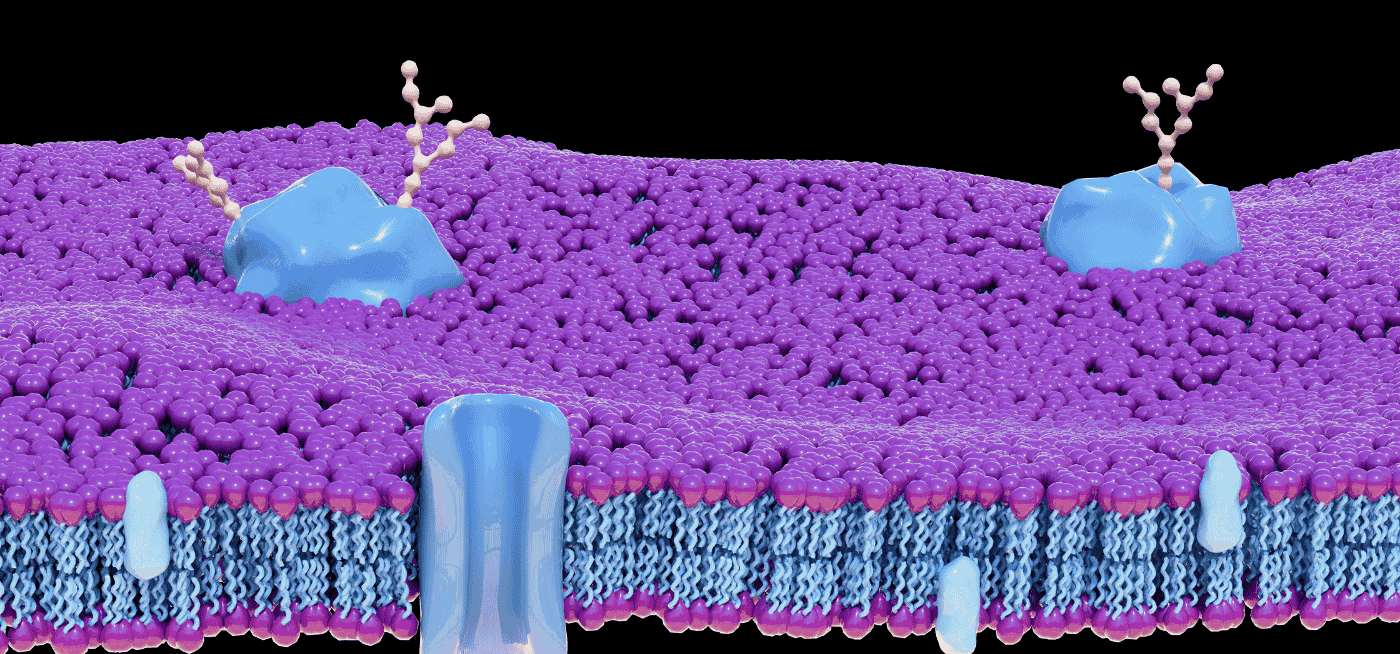
Extracellular vesicles are a heterogeneous population of lipid bilayer-enclosed particles that are released from cells into the extracellular space and biological fluids including blood, urine, saliva, and cerebrospinal fluid. They are broadly classified into three main subtypes based on their biogenesis:
1.1 Exosomes
Exosomes (30–150 nm) originate from the endosomal pathway. Multi-vesicular bodies (MVBs) fuse with the plasma membrane and release intraluminal vesicles into the extracellular environment. They are enriched in tetraspanins (CD9, CD63, CD81), heat shock proteins, and endosomal markers like TSG101 and ALIX.
1.2 Microvesicles
Microvesicles (100–1000 nm) are shed directly from the plasma membrane through outward budding. They carry cytoskeletal proteins, integrins, and surface receptors that reflect the membrane composition of their parent cells.
1.3 Apoptotic Bodies
Apoptotic bodies (1–5 µm) are released during programmed cell death and contain fragmented nuclear material, organelles, and cytoplasmic components. Their cargo provides insight into cell death pathways in disease contexts.
Importantly, EV biogenesis is not random — it is tightly regulated and responsive to cellular stress, hypoxia, inflammation, and oncogenic transformation. This makes EV composition a dynamic readout of cellular biology. Key reference: Théry et al. (2018), MISEV2018 guidelines.
2. EV Profiling: Core Methodologies
Effective EV profiling requires robust isolation, characterization, and cargo analysis strategies. The field has evolved from basic ultracentrifugation to highly sophisticated multi-omics approaches.
2.1 EV Isolation Techniques
- Ultracentrifugation (UC): Gold-standard method for pelleting EVs by size and density.
- Size Exclusion Chromatography (SEC): Preserves EV integrity and removes soluble contaminants.
- Density Gradient Ultracentrifugation: Separates EV subpopulations by buoyant density.
- Polymer Precipitation: Simple and scalable (e.g., ExoQuick, Total Exosome Isolation).
- Microfluidic and Immunoaffinity Capture: Enables high-purity, marker-specific isolation.
2.2 Characterization Platforms
Nanoparticle tracking analysis (NTA) — widely used via platforms like the NanoSight (Malvern Panalytical) — quantifies EV size distribution and concentration. Transmission electron microscopy (TEM) and cryo-EM provide morphological visualization, while flow cytometry enables high-throughput, multi-parameter single-EV analysis.
2.3 Cargo Profiling: Multi-Omics Approaches
- Proteomics: LC-MS/MS-based mass spectrometry identifies the protein cargo of EVs with high sensitivity and depth.
- Transcriptomics: Small RNA sequencing reveals miRNA, lncRNA, and mRNA profiles within EVs.
- Lipidomics: Lipid profiling characterizes the EV membrane and its bioactive lipid cargo.
- Metabolomics: Detects small molecules that reflect metabolic rewiring in disease.
Public EV proteomics and genomics data can be accessed through the EVpedia and Vesiclepedia databases, which catalog thousands of EV-associated proteins and RNAs across cell types and species.
3. EV Profiling in Cancer Biology
Cancer is one of the most extensively studied areas of EV research. Tumor-derived EVs (TDEs) play multifaceted roles in cancer progression, metastasis, and therapy resistance.
3.1 Tumor Microenvironment Remodeling
TDEs educate stromal cells — including fibroblasts, endothelial cells, and immune cells — to create a pro-tumorigenic microenvironment. For example, EVs secreted by pancreatic cancer cells carry TGF-β and fibronectin, which promote fibroblast activation and suppress anti-tumor immunity.
3.2 Pre-Metastatic Niche Formation
Landmark research by Hoshino et al. (2015), published in Nature, demonstrated that tumor-derived exosomes expressing specific integrins preferentially fuse with cells at future metastatic sites, establishing organ-specific pre-metastatic niches. This EV-mediated ‘address labeling’ has opened new avenues for predicting and intercepting metastasis.
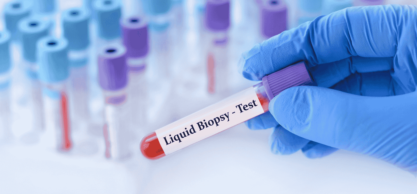
3.3 Liquid Biopsy and Biomarker Discovery
EV profiling is powering a new generation of liquid biopsies. Unlike free circulating tumor DNA (ctDNA), EVs protect their nucleic acid cargo from degradation, offering a stable window into tumor genetics. Proteomic profiling of plasma-derived EVs has identified cancer-specific signatures for breast, lung, colorectal, and ovarian cancers.
Clinical-grade EV-based liquid biopsy platforms are under active development, with companies like Exosome Diagnostics leading efforts to commercialize EV RNA diagnostics for prostate cancer detection.
4. EV Profiling in Neurological Disease
The central nervous system (CNS) poses unique challenges for disease monitoring because the blood-brain barrier restricts access to brain tissue. EVs derived from neurons, astrocytes, and microglia can cross the blood-brain barrier and enter peripheral circulation, making them invaluable for non-invasive CNS disease profiling.
4.1 Alzheimer’s Disease
Neuronal-derived exosomes in plasma carry elevated levels of amyloid-β42, total tau, and phosphorylated tau (p-T181-tau) years before clinical symptom onset. EV profiling thus offers a window into Alzheimer’s pathology that predates conventional biomarker changes in cerebrospinal fluid.
4.2 Parkinson’s Disease
α-Synuclein, a protein that aggregates in Parkinson’s disease, is packaged into EVs and propagated between cells in a prion-like fashion. EV-mediated α-synuclein transfer promotes neuroinflammation and dopaminergic neurodegeneration. Profiling EVs for α-synuclein oligomers may enable early Parkinson’s diagnosis.
4.3 Traumatic Brain Injury and Neuroinflammation
Following traumatic brain injury (TBI), EVs released by damaged neurons carry GFAP, UCH-L1, and neurofilament light chain — proteins with established roles as TBI biomarkers. Real-time EV profiling from blood samples could monitor neurological injury severity and recovery trajectory.
5. EVs in Cardiovascular Disease Biology
Cardiovascular disease (CVD) remains the leading cause of global mortality. EVs derived from cardiomyocytes, endothelial cells, and platelets circulate in blood and carry disease-relevant molecular signatures.
5.1 Myocardial Infarction
During myocardial infarction, ischemic cardiomyocytes release EVs enriched in cardiac troponins, heat shock proteins, and cardioprotective miRNAs (e.g., miR-21, miR-1). These EVs simultaneously reflect cardiac damage and activate downstream cardioprotective signaling in neighboring cells.
5.2 Heart Failure
In heart failure, EV profiling reveals upregulation of pro-fibrotic proteins and inflammatory mediators such as TGF-β and TNF-α. Cardiac fibroblast-derived EVs propagate fibrotic signaling through miR-21-5p, contributing to pathological cardiac remodeling.
5.3 Atherosclerosis
Endothelial cell-derived EVs in atherosclerosis carry intercellular adhesion molecule-1 (ICAM-1) and tissue factor, promoting inflammatory monocyte recruitment and thrombus formation. For a comprehensive review, see Loyer et al. (2018) in Circulation Research.
6. EV Profiling in Infectious Disease and Immune Biology
Pathogens and the immune system engage in a dynamic dialogue mediated in part by EVs. Both host cells and pathogens exploit EV biology to manipulate immunity.
6.1 Viral Infections
Viruses like HIV, SARS-CoV-2, and Epstein-Barr virus (EBV) hijack EV biogenesis pathways to package viral proteins, nucleic acids, and immune-evasion factors. EV profiling in COVID-19 patients revealed enrichment of viral spike protein fragments and host immune dysregulation markers, illuminating disease severity mechanisms.
6.2 Bacterial Infections and Sepsis
Bacterial outer membrane vesicles (OMVs) — functionally analogous to eukaryotic EVs — deliver virulence factors, lipopolysaccharide (LPS), and antibiotic resistance genes to host cells. In sepsis, profiling circulating EVs provides insights into systemic inflammatory cascades and organ dysfunction pathways.
6.3 Immunomodulation
Immune cell-derived EVs, particularly those from dendritic cells, T cells, and NK cells, regulate adaptive and innate immunity. Dendritic cell exosomes presenting MHC-peptide complexes can activate antigen-specific T cells, inspiring therapeutic EV platforms for cancer immunotherapy and vaccination.
7. EV Profiling in Metabolic and Fibrotic Diseases
7.1 Type 2 Diabetes and Obesity
Adipose tissue-derived EVs in obese individuals carry inflammatory miRNAs and lipid species that induce insulin resistance in recipient liver and muscle cells. EV profiling in diabetic patients reveals distinct protein and miRNA signatures associated with beta-cell dysfunction and systemic metabolic dysregulation.
7.2 Non-Alcoholic Fatty Liver Disease (NAFLD)
Hepatocyte-derived EVs are elevated in NAFLD and non-alcoholic steatohepatitis (NASH), carrying ceramide, TRAIL, and miR-192-5p that activate hepatic stellate cells and Kupffer cells to drive fibrosis and inflammation. EV profiling may enable non-invasive liver disease staging, potentially replacing liver biopsy.
7.3 Kidney Disease
Urinary EV profiling offers a non-invasive window into kidney pathology. In chronic kidney disease (CKD) and diabetic nephropathy, urinary EVs carry podocyte-specific proteins (nephrin, podocin) and inflammatory cytokines reflecting glomerular damage. Longitudinal EV monitoring may track disease progression and treatment response.
8. Technological Advances Accelerating EV Profiling
Several technological breakthroughs are dramatically increasing the depth, resolution, and clinical applicability of EV profiling.
8.1 Single-EV Analysis
Conventional bulk EV analysis masks the heterogeneity of individual EV populations. Emerging single-EV technologies — including super-resolution microscopy, single-particle interferometric reflectance imaging sensing (SP-IRIS), and nano-flow cytometry — enable molecule-by-molecule characterization of individual EVs. The ExoView platform (NanoView Biosciences) uses multiplexed antibody chips to simultaneously profile EV surface protein co-expression at single-particle resolution.
8.2 Artificial Intelligence and Machine Learning
Machine learning algorithms are being deployed to decode complex EV multi-omics datasets. AI-driven integration of EV proteomic, transcriptomic, and lipidomic data enables discovery of disease-specific EV fingerprints that would be impossible to detect with conventional statistical methods. Deep learning models trained on EV MS data are achieving high accuracy in cancer type classification.
8.3 Microfluidic EV Chips
Lab-on-chip microfluidic devices can isolate and profile EVs directly from small volumes of clinical samples in under an hour. These point-of-care platforms bring EV diagnostics closer to clinical reality, potentially enabling rapid disease monitoring at the bedside.
8.4 Spatial Transcriptomics of EV Biogenesis
Coupling spatial transcriptomics with EV cargo analysis allows researchers to map EV biogenesis and release within intact tissue architecture, revealing how disease microenvironments shape EV signaling networks.
9. EV Profiling for Therapeutic Development
Beyond diagnostics, EV profiling is catalyzing a new class of EV-based therapeutics.
9.1 Natural EVs as Therapeutic Agents
Mesenchymal stem cell (MSC)-derived EVs exhibit potent anti-inflammatory, pro-angiogenic, and regenerative properties. EV profiling has helped identify the key miRNAs and proteins responsible for these effects, enabling rational optimization of MSC-EV therapeutics for heart failure, acute lung injury, and graft-versus-host disease.
9.2 Engineered EVs as Drug Delivery Vehicles
EVs can be engineered to display targeting ligands and carry therapeutic payloads — including small molecules, siRNA, CRISPR-Cas9 complexes, and chemotherapy drugs. EV profiling guides the selection of optimal parental cell types, loading strategies, and surface modifications to maximize efficacy and minimize off-target effects.
9.3 EV Vaccines
Bacterial OMV-based vaccines are already clinically approved, including vaccines against Neisseria meningitidis. Eukaryotic EV vaccines loaded with tumor antigens or viral proteins are in preclinical and early clinical development. A landmark 2021 trial published in Nature Communications demonstrated EV-based cancer vaccine efficacy in mouse models.
10. Challenges and Standardization in EV Profiling
Despite extraordinary progress, several challenges must be addressed before EV profiling achieves widespread clinical adoption.
10.1 Pre-analytical Variables
EV composition is exquisitely sensitive to sample collection, processing, and storage conditions. Anticoagulant choice, freeze-thaw cycles, storage temperature, and centrifugation parameters all profoundly affect EV yield and cargo profiles, complicating cross-study comparisons.
10.2 EV Heterogeneity and Subtype Resolution
Current bulk profiling methods cannot resolve functional EV subpopulations. Since different EV subtypes carry distinct biological messages, failure to distinguish them limits mechanistic insight. Advances in single-EV analysis and subpopulation-specific isolation are beginning to address this challenge.
10.3 Regulatory and Translational Hurdles
EV-based diagnostics and therapeutics face complex regulatory pathways. The FDA has issued guidance on EV characterization for therapeutic applications. Bodies including the ISEV and the EV-TRACK consortium are working to establish community-wide reporting standards to support regulatory submissions.
Conclusion: EV Profiling as a Pillar of Disease Biology
Extracellular vesicle profiling has fundamentally reshaped our understanding of disease biology. By providing a molecular snapshot of cellular communication networks across cancer, neurodegeneration, cardiovascular disease, infection, and metabolism, EV profiling bridges mechanistic discovery and clinical translation.
As isolation, characterization, and analytical technologies continue to advance, EV profiling is poised to become a cornerstone of precision medicine — enabling earlier disease detection, more accurate prognosis, and rationally designed therapies that harness the body’s own intercellular messaging system.
The field’s future lies in the integration of multi-omics EV data with artificial intelligence, single-EV resolution technologies, and standardized clinical protocols — a convergence that promises to unlock the full diagnostic and therapeutic potential of extracellular vesicles.
References
[2] Hoshino A, et al. (2015). Tumour exosome integrins determine organotropic metastasis. Nature.
[15] Vesiclepedia — Comprehensive EV Molecular Cargo Database.