

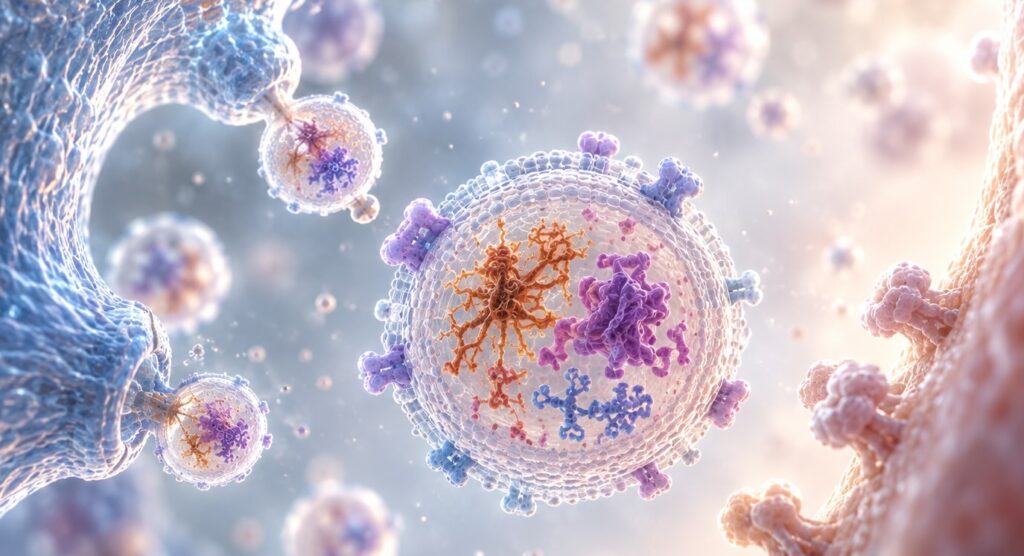
Most of the field treats a vesicle as a container — something that happens to hold tau, or α-synuclein, or TDP-43, and can therefore be sampled. That framing is useful and incomplete. Over the past decade, work across Alzheimer’s, Parkinson’s and ALS models has placed vesicles inside the pathology itself: as the route misfolded protein takes between cells, as the channel through which activated glia reach neurons, and as a pressure valve that opens when degradation fails. The measurement case rests on that biology.
What Is the Role of Extracellular Vesicles in Neurodegeneration?
Extracellular vesicles in neurodegeneration act as a transport system, not a byproduct. They carry misfolded tau, α-synuclein and TDP-43 between cells, propagating pathology along anatomical circuits. They also mediate glial-to-neuronal signaling that drives synaptic dysfunction. Because a vesicle’s surface reflects the cell that released it, its cargo is an interpretable readout of a specific cellular process.
Key takeaways
- Vesicular transfer is an established mechanism of protein spreading in tau, α-synuclein and TDP-43 pathology — demonstrated in vivo, not just in culture.
- Microglial and astrocytic vesicles are active effectors of synaptic and inflammatory dysfunction, not inert reporters of it.
- Vesicle secretion rises when autophagy-lysosomal degradation fails, which means secretion rate itself carries information about cellular state.
- Because cargo composition is inherited from the parent cell, cell of origin — not analyte identity — determines whether a plasma measurement is interpretable.
- Total vesicle counts answer almost nothing. Surface composition and subpopulation structure carry the signal.
Vesicles Are a Transport Route for Misfolded Protein
The case for extracellular vesicles in neurodegeneration begins with a mechanical question the prion-like framing raises but does not answer. Pathology seeded in one region spreads along connected circuits — pathology seeded in one region and spreading along connected circuits. What actually moves between cells? Free protein in the interstitial space is one answer. Vesicular transport is another, and it has accumulated the stronger in vivo evidence.
Tau
The clearest early demonstration came from a mouse model engineered for rapid tau spreading from entorhinal cortex to dentate gyrus. Depleting microglia suppressed that spread substantially, and inhibiting vesicle synthesis reduced it both in culture and in the animal — implicating microglia as a vehicle rather than only a cleanup crew [2]. A later study took vesicles isolated directly from post-mortem human Alzheimer’s brain tissue and injected them into mouse brain, where they propagated tau pathology into interneurons more efficiently than vesicles from control donors [3]. The material is transmissible, and its potency tracks with the disease state of its source.
α-Synuclein
Parkinson’s shows the same architecture. Microglia exposed to α-synuclein preformed fibrils release vesicles that transmit pathology to neurons; injected into mouse striatum, those vesicles produced phosphorylated α-synuclein across regions consistent with neuronal connectivity, along with time-dependent nigrostriatal degeneration [4]. Depleting microglia in vivo sharply reduced transmission. The same work found α-synuclein oligomers in microglia-derived vesicles recovered from patient CSF, which closes some of the gap between the model and the patient.
Mechanistically, the trigger runs through degradation. Preformed fibrils impaired autophagic flux in activated microglia by upregulating PELI1, which in turn drove loss of the lysosomal membrane protein LAMP2 [4]. A cell that cannot degrade what it has taken up exports it instead — and in doing so hands the problem to its neighbours. The same logic recurs across indications, which is part of why it deserves attention: this is not a Parkinson’s-specific quirk but a general failure mode of cells under proteostatic load.
TDP-43 — and a complication worth keeping
ALS and FTLD introduce a tension the field has not fully resolved. TDP-43 is secreted in vesicles from neurons but not from astrocytes or microglia, and vesicular TDP-43 species are elevated in ALS brain [5]. Vesicles from ALS brain caused cytoplasmic TDP-43 redistribution in recipient cells, consistent with propagation. But blocking vesicle secretion made things worse: aggregates accumulated intracellularly, and a TDP-43 mutant mouse treated with a secretion inhibitor deteriorated [5]. Secretion is doing two jobs at once — spreading pathology and relieving the cell that produced it.
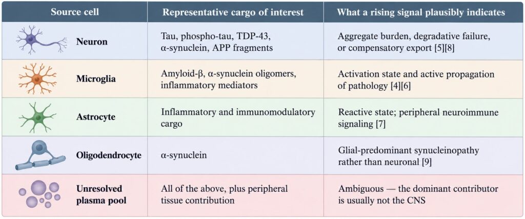
That ambiguity is not a reason to discount vesicles. It is a reason to be precise about what a rising vesicular signal means, which depends entirely on knowing which cell released the vesicle.
Glial Vesicles Are Effectors, Not Bystanders
Neuroinflammation is usually described in terms of cytokines. Vesicles belong in that description. Amyloid-β released by microglia in association with large vesicles alters dendritic spine morphology at the point of neuronal contact and impairs long-term potentiation in vivo [6]. The deficit was observed in entorhinal cortex one hour after injection and had reached the dentate gyrus by 24 hours — a measurable spread of functional impairment, not just of pathological protein.
There is also a feed-forward quality to this signaling that single-timepoint thinking tends to miss. Exposure to amyloid-β oligomers raises ceramide levels in microglia through acid sphingomyelinase activation, prompting release of proinflammatory factors; those factors activate astrocytes, which release ceramide-enriched vesicles of their own; microglia take those up, and the inflammatory response amplifies [1]. A self-sustaining loop of this kind means a vesicle measurement at any one moment is sampling a process with its own momentum, not a static state.
Astrocytic vesicles carry a comparable weight. They participate in CNS neuroimmune responses and can act well beyond the parenchyma, with vesicles emanating from the brain into circulation and influencing cells in peripheral immune organs [7]. The implication for anyone building a blood-based assay is direct: a plasma vesicle pool is not a passive spillover of brain contents. Some of it is signaling that was always meant to leave.
When Degradation Fails, Secretion Rises
Endolysosomal and autophagic dysfunction sits near the centre of neurodegenerative pathology, and it connects to vesicle biology in a mechanistically specific way. Disrupting neuronal Vps34 function — the class III PI 3-kinase that produces phosphatidylinositol-3-phosphate — impairs autophagy and lysosomal degradation, damages endolysosomal membranes, and promotes the release of vesicles loaded with undigested lysosomal substrates, including amyloid precursor protein C-terminal fragments and distinctive lipid species [8].
Read that alongside the TDP-43 finding and a pattern emerges. Cells under degradative stress export what they cannot break down. Vesicle release is partly a function of how badly the degradation machinery is coping. That makes secretion rate and cargo composition a joint readout of cellular state — but only if the cell of origin is known, because the same total-vesicle measurement in plasma aggregates output from platelets, endothelium, erythrocytes, hepatocytes and every other tissue in the body.
How CNS Vesicles Reach the Bloodstream
None of this would matter for blood-based work if vesicles stayed inside the skull. They do not. Vesicles traverse the blood–brain barrier in both directions, and that property is the reason a peripheral draw can report on a central process at all [1]. It is also the property that makes vesicle-based drug delivery an active area of development, for the same physical reasons.
But traversal cuts both ways. A vesicle that can leave the brain enters a compartment in which it is heavily outnumbered. Circulating vesicles derive from platelets, erythrocytes, endothelium, liver, adipose tissue, skeletal muscle and immune cells, all contributing to the same pool at far higher abundance than the CNS does [10]. The brain’s contribution is real, informative, and a small minority of the total.
This is the structural reason why sensitivity improvements alone have not resolved the blood-based CNS biomarker problem. Pushing a detection limit lower measures the mixed pool more precisely. It does not change what is in the pool. The constraint is compositional, and compositional constraints are addressed upstream — at enrichment — rather than downstream at detection.
What the Biology Demands of Measurement
Here is where mechanism and assay design meet. Vesicles reflect the phenotypic state of the cell that released them, which is exactly what makes them attractive for liquid biopsy — and the heterogeneity of circulating vesicles in surface composition, biogenesis and cellular origin is exactly what makes selective enrichment difficult [10]. Both statements are true simultaneously, and the second one governs whether the first is usable.
The most instructive demonstration is a study that isolated vesicles from patient serum and plasma by immunoaffinity capture against cell-type-restricted surface epitopes — separating putative neuronal from putative oligodendroglial populations — and measured α-synuclein in each [9]. Concentrations were lowest in controls and highest in multiple system atrophy relative to Parkinson’s. But the sharpest discriminator was not either concentration on its own. It was the ratio between the two populations. A model combining that ratio with α-synuclein concentration and total vesicle concentration separated Parkinson’s from MSA with an AUC of 0.902, corresponding to 89.8% sensitivity and 86.0% specificity in an independent validation cohort [9].
That result is worth sitting with. The same analyte, measured in the same blood draw, meant different things depending on which cell released the vesicle carrying it. Provenance was not a technical footnote. It was the discriminating variable.
Figure 1. What vesicle origin changes about interpretation
A note on nomenclature, since the terms are used loosely. “Exosome” refers specifically to vesicles of endosomal origin, released when a multivesicular body fuses with the plasma membrane. Because origin is difficult to establish for vesicles recovered from a biofluid, current consensus guidance recommends the operational term “extracellular vesicle” unless biogenesis has been demonstrated [11]. The same guidance emphasises characterising vesicles with complementary methods rather than relying on a single measure. We use “extracellular vesicle” throughout for that reason.
Where This Is Heading
Three problems are open, and none of them are small.
The first is causality. Most propagation evidence comes from models in which vesicles are concentrated, injected, or blocked pharmacologically. Those manipulations establish sufficiency well and necessity less well. Whether vesicular transfer is the dominant route of spread in sporadic human disease, or one route among several, is not settled [1].
The second is resolution. Bulk measurement across a vesicle population obscures the very heterogeneity that carries the signal, and single-vesicle technologies are advancing quickly to address it — with standardisation named as the critical bottleneck for clinical translation [12]. A rare subpopulation with a distinctive surface signature is invisible to any assay that reports a population average.
The third is pre-analytical. Vesicle secretion responds to cellular stress, which means it also responds to how blood is collected, handled and stored. Time to centrifugation, anticoagulant choice, centrifugation protocol, freeze–thaw history and even the degree of haemolysis all shift what ends up in the measured pool — and platelet-derived vesicles in particular can be generated after the draw, by handling rather than by biology. Signals that move with processing conditions will not survive contact with a multi-site study, regardless of how elegant the underlying mechanism is. Consensus guidance now treats pre-analytical reporting as a minimum requirement rather than a methods-section courtesy [11].
What has changed is the starting assumption. A decade ago, extracellular vesicles in neurodegeneration were interesting mainly because they were measurable. They are now understood as participants — carrying pathology between cells, mediating glial signaling, and venting what damaged cells cannot degrade. That reframing raises the bar for measurement rather than lowering it. If a vesicle’s cargo reflects a specific cellular process, then the assay has to be able to say which cell, and which process. Everything else is a number without a subject.
That requirement — multiplex surface protein profiling of plasma vesicle subpopulations, without a prior isolation step — is what the LuminEV Research Kit was designed to meet. It is one approach to a problem the biology has made unavoidable, and the problem will outlast any particular solution to it.
References
- Chen J, Tian C, Xiong X, Yang Y, Zhang J. Extracellular vesicles: new horizons in neurodegeneration. eBioMedicine. 2025;113:105605. doi:10.1016/j.ebiom.2025.105605
- Asai H, Ikezu S, Tsunoda S, et al. Depletion of microglia and inhibition of exosome synthesis halt tau propagation. Nat Neurosci. 2015;18(11):1584–1593. doi:10.1038/nn.4132
- Ruan Z, Pathak D, Venkatesan Kalavai S, et al. Alzheimer’s disease brain-derived extracellular vesicles spread tau pathology in interneurons. Brain. 2021;144(1):288–309. doi:10.1093/brain/awaa376
- Guo M, Wang J, Zhao Y, et al. Microglial exosomes facilitate α-synuclein transmission in Parkinson’s disease. Brain. 2020;143(5):1476–1497. doi:10.1093/brain/awaa090
- Iguchi Y, Eid L, Parent M, et al. Exosome secretion is a key pathway for clearance of pathological TDP-43. Brain. 2016;139(12):3187–3201. doi:10.1093/brain/aww237
- Gabrielli M, Prada I, Joshi P, et al. Microglial large extracellular vesicles propagate early synaptic dysfunction in Alzheimer’s disease. Brain. 2022;145(8):2849–2868. doi:10.1093/brain/awac083
- Sutter PA, Lavoie ER, Lombardo ET, Pinter MK, Crocker SJ. Emerging role of astrocyte-derived extracellular vesicles as active participants in CNS neuroimmune responses. Immunol Invest. 2024;53(1):26–39. doi:10.1080/08820139.2023.2281621
- Miranda AM, Lasiecka ZM, Xu Y, et al. Neuronal lysosomal dysfunction releases exosomes harboring APP C-terminal fragments and unique lipid signatures. Nat Commun. 2018;9(1):291. doi:10.1038/s41467-017-02533-w
- Dutta S, Hornung S, Kruayatidee A, et al. α-Synuclein in blood exosomes immunoprecipitated using neuronal and oligodendroglial markers distinguishes Parkinson’s disease from multiple system atrophy. Acta Neuropathol. 2021;142(3):495–511. doi:10.1007/s00401-021-02324-0
- Pierri B, Eitan E, Witwer KW, Re DB, Baccarelli AA, Wu H. Tissue-specific extracellular vesicles enriched from circulation: exploring the liquid biopsy perspective. J Extracell Biol. 2026;5(2):e70106. doi:10.1002/jex2.70106
- Welsh JA, Goberdhan DCI, O’Driscoll L, et al. Minimal information for studies of extracellular vesicles (MISEV2023): from basic to advanced approaches. J Extracell Vesicles. 2024;13(2):e12404. doi:10.1002/jev2.12404
- Zhang Y, Meng X, Greening DW, et al. Unveiling heterogeneity: innovations and challenges in single-vesicle analysis for clinical translation. J Extracell Vesicles. 2025;14(12):e70209. doi:10.1002/jev2.70209