

Synapse loss begins years before a patient forgets a name, and it tracks cognitive decline more closely than plaques or tangles do [1]. For most of the field’s history, counting synapses meant a microscope and a post-mortem brain. SV2A PET now measures synaptic density in living patients, but only at centers with the tracer [2]. CSF offers synaptic proteins at the cost of a lumbar puncture. A third window is opening in blood, and it runs through the vesicles neurons release.
What are synaptic extracellular vesicles?
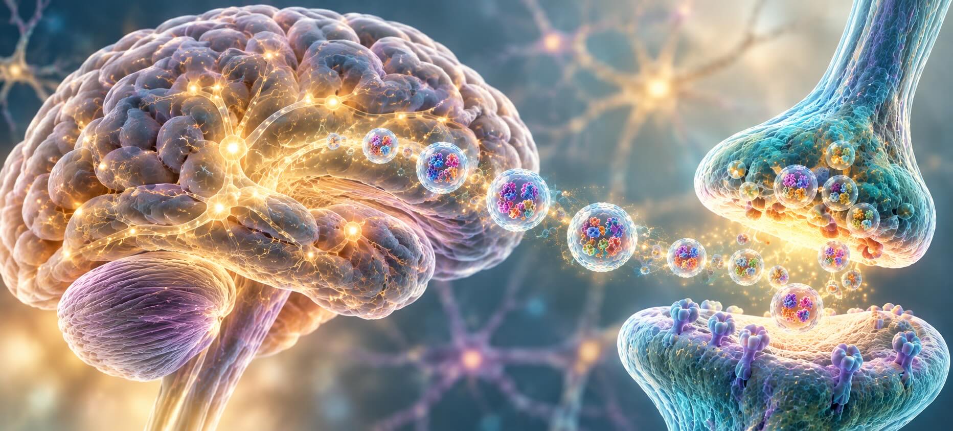
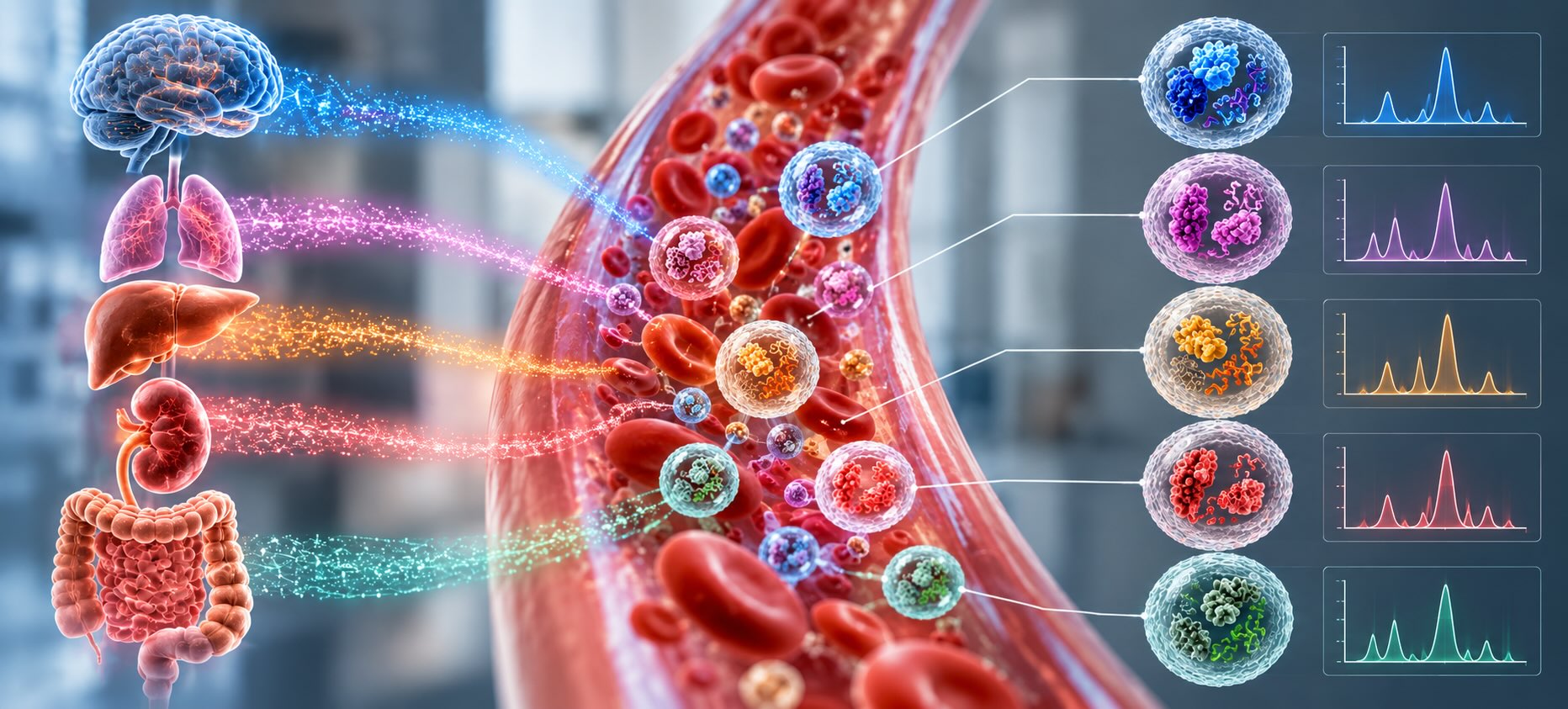
Synaptic extracellular vesicles are membrane-bound particles released by neurons that carry synaptic proteins, RNA and lipids between cells. They take part in activity-dependent signaling and can reach peripheral blood, where their cargo reflects synaptic health. They are distinct from synaptic vesicles, which store neurotransmitter inside the nerve terminal.
The naming overlap causes real confusion, so it is worth being precise. A synaptic vesicle is a roughly 40 nm organelle that stays inside the presynaptic terminal and fuses with the plasma membrane to release glutamate, GABA or another transmitter. It never leaves the cell as an intact particle. An extracellular vesicle (EV) does. MISEV2023 recommends “extracellular vesicle” as the umbrella term and reserves “exosome” for vesicles shown to originate in the endosomal pathway, released when a multivesicular body fuses with the plasma membrane [3]. Because most plasma studies cannot prove that biogenesis route, this article uses EV throughout.
“Synaptic EV” is best read as an operational term: a neuron-derived vesicle that carries synaptic cargo. Whether that vesicle physically left the cell at the synapse is a separate question, and the answer turns out to matter for how the signal is interpreted.
Key Takeaways
- Neurons load synaptic extracellular vesicles selectively. Their RNA and protein content differs from the parent cell, so composition carries information about cellular state.
- Synaptic proteins in neuron-enriched plasma EVs fall in Alzheimer’s disease and FTD, and in several cohorts the decline appears 5 to 11 years before dementia.
- Presynaptic and postsynaptic proteins do not decline in lockstep. Paired and multiplexed readouts carry information that single proteins miss.
- The largest open question is anatomical: much neuronal EV biogenesis happens in the soma and dendrites, not at axon terminals.
The synapse talks in vesicles
The case for measuring synaptic EVs rests on a simpler observation: vesicles are part of how synapses work, not a waste stream. Three lines of evidence make the point.
Arc: a memory gene that packages RNA
Arc is an activity-regulated gene required for long-term memory storage and several forms of synaptic plasticity. In 2018, Pastuzyn and colleagues showed that Arc protein self-assembles into capsids resembling those of retroviruses, encapsulates RNA, and leaves neurons inside extracellular vesicles. Those vesicles deliver Arc mRNA into recipient cells, where it can undergo activity-dependent translation [4]. Evolutionary analysis traced Arc to a vertebrate lineage of Ty3/gypsy retrotransposons, the same family that gave rise to retroviruses.
A companion study in the same issue of Cell found the fly version, Arc1, binds RNA and traffics across synaptic boutons at the neuromuscular junction, moving from presynaptic neuron to postsynaptic muscle [5]. The two lineages arose independently. Evolution arrived at vesicle-borne RNA transfer across the synapse at least twice, which suggests the function is not incidental.
Neurons tune astrocytes through vesicle microRNA
Most EV signaling data come from culture, where release rates and cargo can differ sharply from tissue. Men and colleagues addressed this with a cell-type-specific reporter mouse that labels the endosomal precursors of neuronal vesicles in vivo [6]. Neuronal vesicles carried a microRNA profile distinct from that of the neurons releasing them, evidence of selective loading. One neuron-specific microRNA, miR-124-3p, was taken up by astrocytes, where it raised levels of GLT1, the dominant glutamate transporter in the brain.
That is a synaptic function by any definition. Astrocytic glutamate clearance sets how long transmitter lingers in the cleft, and here neurons are adjusting it through vesicle cargo. It also means vesicle composition is regulated, not a random sample of cytoplasm. A change in what neurons export is a change in neuronal state.
The same machinery, a different cargo
The pathways that carry Arc capsids and microRNA can also carry misfolded tau, α-synuclein and TDP-43 between cells, a mechanism covered in detail in our companion article on vesicles as active participants in neurodegeneration. The focus here is narrower: what happens to the synaptic cargo itself when synapses begin to fail.
When synapses fail, the cargo changes
If vesicle content reflects neuronal state, synapse loss should leave a mark on the cargo of synaptic extracellular vesicles. The plasma data now support that prediction across several independent cohorts.
An early study enriched neuron-derived vesicles from plasma by immunoaffinity capture against neuron-restricted surface epitopes, then quantified six synaptic proteins normalized to vesicle quantity. The cross-sectional arm included 12 patients with Alzheimer’s disease, 16 with FTD and 28 controls [7]. Synaptophysin, synaptopodin, synaptotagmin-2 and neurogranin were significantly lower in both dementias. A growth-associated presynaptic protein and synapsin 1 fell only in Alzheimer’s disease. In the longitudinal arm, synaptotagmin, synaptophysin and neurogranin were already reduced years before dementia. Several synaptic proteins correlated with cognitive scores, whereas vesicle Aβ42 and p-tau181 did not.
Jia and colleagues then asked whether vesicle synaptic proteins could identify preclinical Alzheimer’s disease in cognitively normal people [8]. Their design had an important feature: the blood vesicle measurements were validated against concomitant CSF findings in the same participants. A combination of four proteins detected preclinical disease 5 to 7 years before cognitive impairment. The four were the growth-associated presynaptic protein, neurogranin, SNAP25 and synaptotagmin 1.
Brain tissue points the same way. Muraoka and colleagues profiled vesicles isolated directly from the cortical gray matter of 20 Alzheimer’s and 18 control brains [9]. Control vesicles were rich in neuron-specific proteins, while Alzheimer’s vesicles carried more glia-specific proteins. A machine-learning signature separated the two groups with 88% accuracy in held-out test sets. The vesicle pool of a diseased cortex has a different cellular makeup, not just different levels of individual proteins.
A 2025 systematic review pooled 34 studies and 5,601 participants and reached a consistent conclusion. Synaptic proteins in blood neural-derived vesicles change early in the clinical course, and vesicle markers predicted Alzheimer’s disease across horizons of 1 to 10 years [10]. The review also documents the field’s weak points: small cohorts, varied isolation methods, and few replications of any single assay.
A lower synaptic signal per vesicle can mean three different things. There may be fewer synapses. There may be fewer neuronal vesicles in the enriched fraction. Or loading may have changed while synapse number held steady. The studies above normalize to vesicle quantity for exactly this reason, but normalization only controls for the second explanation. Separating synapse loss from altered loading requires measuring more than one protein at a time.
Presynaptic, postsynaptic: why one marker isn’t enough
A synapse has two sides built from different proteins, and they do not fail on the same schedule. That asymmetry is where most of the diagnostic information lives.
Goetzl and colleagues tested it directly by measuring two matched pairs from excitatory synapses in neuron-enriched plasma vesicles [11]. The presynaptic proteins were neuronal pentraxin 2 and neurexin 2α. Their postsynaptic partners were the GluA4-containing AMPA receptor and neuroligin 1. All four were lower in Alzheimer’s dementia (n = 46), but only the two postsynaptic proteins correlated with the extent of cognitive loss. In samples drawn 6 to 11 years before dementia onset, three of the four were already reduced, and neuronal pentraxin 2 was the exception. Partners that work together at the same synapse gave different clinical information and diverged in timing.
The first study showed the same thing between diseases. Some synaptic proteins fell in both Alzheimer’s disease and FTD, while others fell only in Alzheimer’s disease [7]. No single protein distinguished the two conditions. The pattern did.
CSF work in frontotemporal lobar degeneration makes the multiplexing argument quantitatively. Cervantes González and colleagues measured eight synaptic proteins in the antemortem CSF of an autopsy-confirmed cohort [12]. A multimarker panel that included calsyntenin-1 was associated with postmortem TDP-43 burden (r² = 0.69). A second panel discriminated FTLD-tau from FTLD-TDP with an AUC of 0.83. Both are pathological distinctions that no individual protein resolved as well.
Reading synaptic identity off the vesicle surface
Most of the plasma studies above lysed the enriched vesicle fraction and measured the contents in bulk. Tian and colleagues took a different route [13]. They identified NMDAR2A, an NMDA receptor subunit, as a surface marker of CNS-derived plasma vesicles and counted individual vesicles by flow cytometry. Vesicles carrying the marker were reduced in Alzheimer’s disease compared with healthy controls in a discovery cohort and in an independent multicenter validation cohort. Models that combined these vesicles with Alzheimer’s markers carried on the same vesicles reached AUCs of 0.915 and 0.810, respectively.
Several of the most informative synaptic proteins are transmembrane: receptors, neurexins, neuroligins, synaptotagmins. That makes surface profiling a natural fit for this biology, since a receptor subunit on the vesicle membrane identifies the compartment it came from. The practical requirements follow from the evidence above. An assay needs neuron-enriched input, normalization to a general vesicle marker such as the tetraspanins CD9, CD63 and CD81, and enough simultaneous readouts to pair presynaptic with postsynaptic proteins. It also needs to fit within the plasma volume a cohort can spare.
Multiplex bead-based immunoassays such as the LuminEV Research Kit are one way to meet those requirements. They profile several surface proteins on the same vesicle population from a single plasma aliquot, which keeps paired readouts on a shared denominator rather than across separate runs. Whatever the platform, MISEV2023 reporting is the minimum bar for results that other groups can compare [3]. That means documented isolation, characterization and particle normalization. Because LuminEV panels can be customized, a lab can put presynaptic and postsynaptic targets and a tetraspanin normalizer in a single panel design.
What we still don’t know
The evidence that synaptic extracellular vesicles in plasma track disease is consistent. The evidence for how that signal is generated is thinner, and four questions stand out.
Where the vesicles leave the neuron. The reporter mouse study found neuronal vesicle precursors mainly in the soma and dendrites, not in axon terminals [6]. If that holds broadly, presynaptic proteins in plasma vesicles may reflect what the cell body is producing and sorting rather than what the terminal is shedding. Postsynaptic cargo, released from dendrites, may be closer to a direct readout. The distinction shapes how a falling presynaptic signal should be interpreted, and it has not been tested systematically in humans.
How the plasma level is set. A plasma measurement reflects production minus clearance. Transit across the blood-brain barrier, circulating half-life and uptake by peripheral cells are poorly characterized for neuronal vesicles. Any of them could shift with age, inflammation or treatment independently of synapse number.
Which synapses are reporting. Guo and colleagues identified plasma vesicle markers enriched in brain regions vulnerable to Alzheimer’s disease [14]. Combining regional identity with synaptic cargo could separate hippocampal from cortical synapse loss, or excitatory from inhibitory, but that work is only beginning.
Whether the signal moves with treatment. The strongest use case may be pharmacodynamic. The Synaptic Health Endpoints Working Group has argued that synaptoprotective drugs need biomarkers that quantify synapse damage in trials [1]. Plasma vesicles allow sampling as often as a protocol needs. What is missing is longitudinal evidence that vesicle synaptic cargo responds to an intervention on a timescale relevant to trials.
The next phase of research on synaptic extracellular vesicles will look less like biomarker discovery and more like cell biology. The work ahead is to map where neuronal vesicles are born, how their cargo is sorted and how long they circulate. Then the plasma signal has to be anchored to SV2A PET and CSF in the same participants over time. The synapse has been exporting a record of its condition all along. The task now is learning to read it at the resolution the biology offers.
References
- Colom-Cadena M, Spires-Jones T, Zetterberg H, et al. The clinical promise of biomarkers of synapse damage or loss in Alzheimer’s disease. Alzheimers Res Ther. 2020;12(1):21. https://doi.org/10.1186/s13195-020-00588-4
- Chen MK, Mecca AP, Naganawa M, et al. Assessing synaptic density in Alzheimer disease with synaptic vesicle glycoprotein 2A positron emission tomographic imaging. JAMA Neurol. 2018;75(10):1215-1224. https://doi.org/10.1001/jamaneurol.2018.1836
- Welsh JA, Goberdhan DCI, O’Driscoll L, et al. Minimal information for studies of extracellular vesicles (MISEV2023): From basic to advanced approaches. J Extracell Vesicles. 2024;13(2):e12404. https://doi.org/10.1002/jev2.12404
- Pastuzyn ED, Day CE, Kearns RB, et al. The neuronal gene Arc encodes a repurposed retrotransposon Gag protein that mediates intercellular RNA transfer. Cell. 2018;172(1-2):275-288.e18. https://doi.org/10.1016/j.cell.2017.12.024
- Ashley J, Cordy B, Lucia D, Fradkin LG, Budnik V, Thomson T. Retrovirus-like Gag protein Arc1 binds RNA and traffics across synaptic boutons. Cell. 2018;172(1-2):262-274.e11. https://doi.org/10.1016/j.cell.2017.12.022
- Men Y, Yelick J, Jin S, et al. Exosome reporter mice reveal the involvement of exosomes in mediating neuron to astroglia communication in the CNS. Nat Commun. 2019;10(1):4136. https://doi.org/10.1038/s41467-019-11534-w
- Goetzl EJ, Kapogiannis D, Schwartz JB, et al. Decreased synaptic proteins in neuronal exosomes of frontotemporal dementia and Alzheimer’s disease. FASEB J. 2016;30(12):4141-4148. https://doi.org/10.1096/fj.201600816R
- Jia L, Zhu M, Kong C, et al. Blood neuro-exosomal synaptic proteins predict Alzheimer’s disease at the asymptomatic stage. Alzheimers Dement. 2021;17(1):49-60. https://doi.org/10.1002/alz.12166
- Muraoka S, DeLeo AM, Sethi MK, et al. Proteomic and biological profiling of extracellular vesicles from Alzheimer’s disease human brain tissues. Alzheimers Dement. 2020;16(6):896-907. https://doi.org/10.1002/alz.12089
- Pan W, Teng Y, Han X, et al. Value of blood neural cell-derived small extracellular vesicles in the diagnosis and prediction of Alzheimer’s disease: A systematic review. J Prev Alzheimers Dis. 2025;12:100193. https://doi.org/10.1016/j.tjpad.2025.100193
- Goetzl EJ, Abner EL, Jicha GA, Kapogiannis D, Schwartz JB. Declining levels of functionally specialized synaptic proteins in plasma neuronal exosomes with progression of Alzheimer’s disease. FASEB J. 2018;32(2):888-893. https://doi.org/10.1096/fj.201700731R
- Cervantes González A, Irwin DJ, Alcolea D, et al. Multimarker synaptic protein cerebrospinal fluid panels reflect TDP-43 pathology and cognitive performance in a pathological cohort of frontotemporal lobar degeneration. Mol Neurodegener. 2022;17(1):29. https://doi.org/10.1186/s13024-022-00534-y
- Tian C, Stewart T, Hong Z, et al. Blood extracellular vesicles carrying synaptic function- and brain-related proteins as potential biomarkers for Alzheimer’s disease. Alzheimers Dement. 2023;19(3):909-923. https://doi.org/10.1002/alz.12723
- Guo Z, Tian C, Shi Y, et al. Blood-based CNS regionally and neuronally enriched extracellular vesicles carrying pTau217 for Alzheimer’s disease diagnosis and differential diagnosis. Acta Neuropathol Commun. 2024;12(1):38. https://doi.org/10.1186/s40478-024-01727-w