
A decade of extracellular vesicle research settled one question: vesicles in blood carry molecular signal from diseased tissue. It left a harder one open. When a plasma EV measurement changes, which tissue changed? Most studies cannot say, and many cannot reproduce their own numbers with a different isolation method. The next phase of EV liquid biopsy will be decided less by new biomarker candidates than by whether the field can resolve vesicle origin, at scale, with methods that give the same answer twice.
Key Takeaways
- EV liquid biopsy has proven that vesicles carry disease-relevant cargo; its translation has stalled on method heterogeneity and unresolved cell of origin.
- Tissue-resolved EV measurement is the first major shift: in matched human brain tissue and serum, neuronal EV cargo tracked brain content more closely than serum or total EVs.
- Scalable workflows that reduce or remove isolation steps are the second shift, driven by the sample numbers that population studies and trials require.
- Shared standards, reference materials, and fit-for-purpose validation are the third, and they will determine which EV assays reach clinical decisions.
Where Is EV-Based Liquid Biopsy Headed?
EV-based liquid biopsy is moving through three shifts: from measuring bulk vesicle populations to measuring vesicles resolved by tissue of origin, from isolation-heavy workflows to scalable ones suited to large cohorts, and from method diversity to shared standards and reference materials. Together they convert a promising research signal into a reproducible, interpretable clinical measurement.
Each shift answers a specific failure of the first decade. Bulk measurements mix signal from every tissue that sheds vesicles into blood, so a change cannot be attributed. Isolation-heavy workflows limit throughput and introduce method-specific bias, so results do not transfer between laboratories. And without shared reference materials, no one can tell whether two studies disagree because the biology differs or because the measurement does. The sections below take each shift in turn, then look at what it means for teams designing EV studies now.
What the First Decade Delivered, and Where It Stalled
The core premise of EV-based liquid biopsy has held up. Extracellular vesicles are lipid bilayer particles released by essentially all cell types, and their protein, RNA, and lipid cargo reflects the state of the cell that produced them [1]. They are present in blood, urine, cerebrospinal fluid, and saliva, and they protect their cargo from degradation. For a clinician who cannot biopsy a tumor in the leptomeninges or a neuron in the substantia nigra, that combination is the whole point.
What has not held up is comparability. A 2026 systematic review of EV-based liquid biopsy for leptomeningeal metastasis is a representative example. Ten studies met inclusion criteria, and several reported strong discrimination for individual microRNA and protein candidates. Yet heterogeneity in EV isolation techniques, together with small sample sizes, made a meta-analysis unfeasible [2]. The candidates may be real. The literature cannot yet show it, because the studies did not measure the same thing the same way.
The field saw this coming. In 2017, the EV-TRACK consortium launched a crowdsourced knowledgebase to record how EV experiments were performed, arguing that transparent reporting was a precondition for interpreting and replicating results [3]. The International Society for Extracellular Vesicles has updated its minimal information guidelines twice since the first edition in 2014, most recently as MISEV2023, which names separation of EVs from non-vesicular extracellular particles, characterization, and nomenclature as persistent hurdles to clinical application [4] [1]. The problem, in other words, is well described. The next decade is about closing it.
Figure 1. Milestones in standardizing EV-based liquid biopsy research.
| Year | Milestone | What it addressed |
|---|---|---|
| 2014 | First ISEV minimal information guidelines (MISEV2014) | Baseline expectations for EV characterization |
| 2017 | EV-TRACK knowledgebase launched [3] | Transparent reporting of isolation and characterization methods |
| 2018 | MISEV2018 [4] | Expanded characterization and reporting criteria |
| 2019 | Recombinant EVs described as biological reference material [5] | Measuring recovery and technical variability across methods |
| 2024 | MISEV2023 [1] | Updated guidance covering biofluids, tissues, and advanced approaches |
| 2025 | FDA guidance on bioanalytical method validation for biomarkers [6] | Fit-for-purpose validation expectations for biomarker assays |
Standards milestones for extracellular vesicle biomarkers. Sources as cited; MISEV2014 is summarized from MISEV2018 and MISEV2023.
Shift One: From Total EVs to Tissue-Resolved EVs
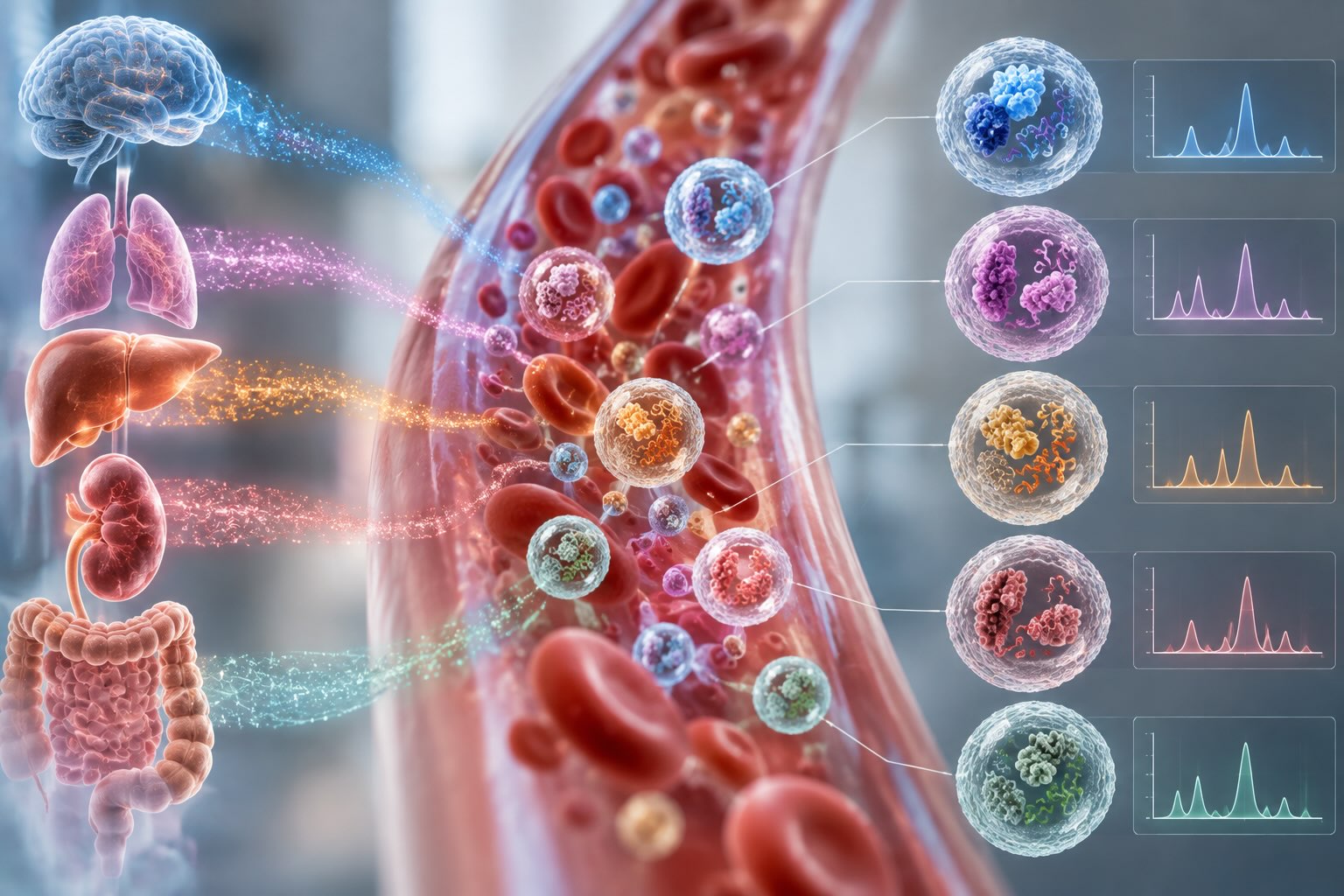
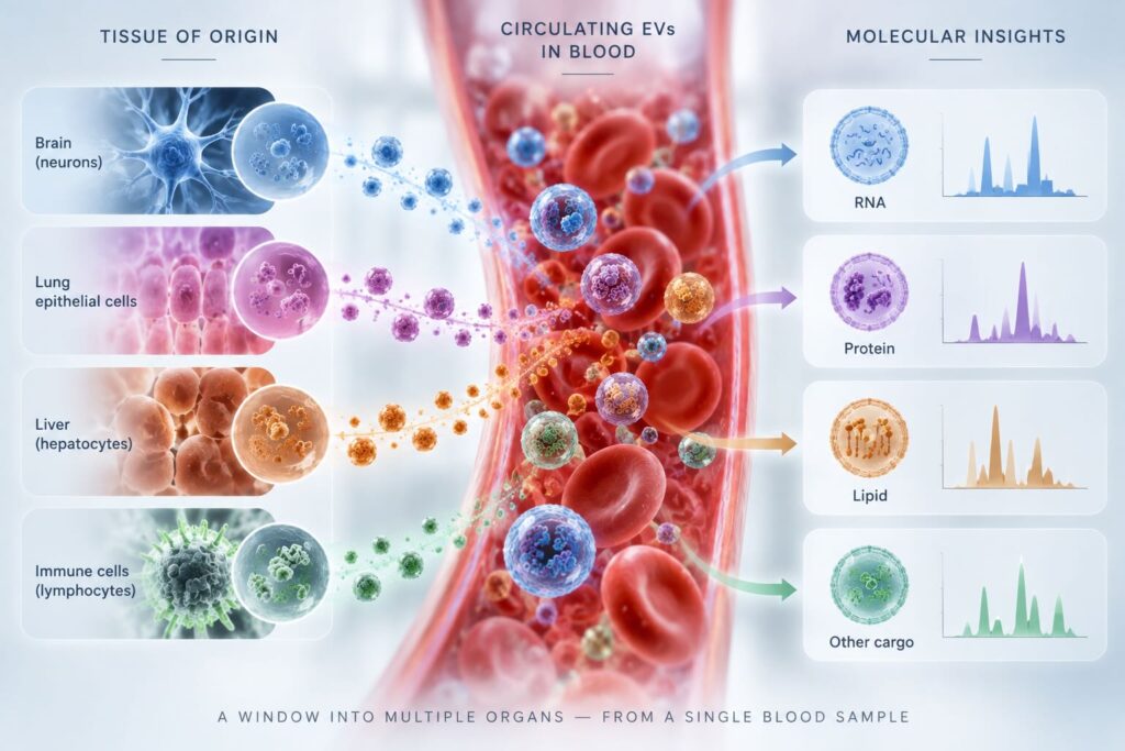
Most circulating EVs come from blood cells, platelets, and endothelium. Transcriptomic deconvolution studies estimate that solid tissues may contribute as little as 0.2% of the circulating pool, and lipoprotein particles of similar size and density outnumber EVs by several orders of magnitude [7]. A measurement of total plasma EVs is therefore a weighted average across every tissue in the body, with the weights set by the tissues that shed the most. For a disease confined to one organ, most of the signal in a total EV measurement comes from somewhere else.
Tissue-specific extracellular vesicles address this by enriching the vesicles from one source before measuring their cargo. The usual approach is immunoaffinity capture against surface proteins expressed predominantly by the tissue of interest. A 2025 review of detection and isolation strategies for tissue-specific EVs from blood described the principle and the practical challenges: suitable markers are limited and can cross-react, surface expression is heterogeneous enough that a significant share of vesicles from the target tissue may lack the capture marker altogether, and selectivity has to be validated with supplementary markers of the target cell type and exclusion of markers from other tissues [7]. A 2026 review in the Journal of Extracellular Biology, co-authored by NeuroDex scientists with investigators at Columbia, Johns Hopkins, and Harvard, reached the same conclusion from the liquid biopsy side, identifying the lack of standardized methods to enrich and characterize tissue-specific EVs from complex biofluids as the major current limitation [8].
The central empirical question is whether enriched vesicles actually report on their tissue. For the brain, a direct test is now available. Using matched sets of human brain tissue and serum, investigators at Columbia and Harvard, with NeuroDex co-authors, compared cargo in serum, serum total EVs, and serum neuronal EVs [9]. MicroRNAs highly expressed in brain tissue correlated more strongly with neuronal EV cargo than with serum or total EV cargo. Environmental chemical concentrations in neuronal EVs also tracked brain tissue levels more closely than serum or total EVs did. That is the evidentiary step the field has needed: not that neuronal EVs contain interesting molecules, but that their contents resemble the organ they are supposed to represent..
Figure 2. Bulk versus tissue-resolved EV measurement.
| Property | Total plasma EVs | Tissue-resolved EVs |
|---|---|---|
| Contributing tissues | All tissues that shed vesicles into blood | Predominantly the target tissue |
| Interpretation of a change | Cannot be attributed to one organ | Attributable, within the specificity of enrichment |
| Signal from a small organ | Diluted by high-output tissues | Concentrated before measurement |
| Main validation question | Is the signal vesicular? | Is the signal vesicular, and from the claimed source? |
| Throughput constraint | Isolation method | Isolation plus enrichment |
Illustrative comparison. Tissue resolution trades additional workflow steps for interpretability.
The implications reach beyond neurology. Circulating EVs with putative origins in brain, liver, adipose tissue, intestine, skeletal muscle, heart, lung, and placenta have all been reported, alongside tumor-derived EVs captured with multi-marker panels, and each faces the same validation questions [7]. For oncology, where tumor-derived vesicles are a small fraction of the circulating pool, the logic is identical to the brain case. For readers following the neurodegeneration application specifically, our earlier analysis of what neuron-derived EVs add to GFAP, NfL, and TDP-43 measurement covers the marker-level evidence.
Shift Two: From Isolation-Dependent to Scalable Workflows
The second shift is about numbers. Discovery studies can tolerate slow, multi-step isolation because they run dozens of samples. Validation cohorts, population studies, and clinical trials run hundreds to thousands, often across sites and years. At that scale, every isolation step is a source of sample loss, method-specific bias, and variance between operators, and the heterogeneity that blocked the leptomeningeal meta-analysis becomes a design problem inside a single study.
Two developments are converging on this constraint. The first is multiplexed measurement of EV surface proteins directly in plasma or culture media, without a separate isolation step. In NeuroDex’s own published validation of the LuminEV assay, a bead-based multiplex immunoassay for EV surface proteins, measurement of tetraspanins CD9, CD63, and CD81 in plasma from 70 disease-free donors showed relative abundances of 72%, 16%, and 12%, respectively, with CD63 showing a weak but significant negative correlation with age and slightly lower levels in female samples [10]. The same study detected cell type-associated surface markers for erythrocytes, neurons, and macrophages, and differences in tetraspanin profiles between healthy and diseased donors.
Those numbers matter for the future of the field in a specific way. If the dominant EV tetraspanin in plasma accounts for roughly three-quarters of the signal and the minor ones vary with donor characteristics, then normalizing a biomarker to “total EVs” depends heavily on which surface protein defines the total. Surface profiling at scale turns that from a hidden assumption into a measured variable. The second development is the pairing of that kind of profiling with tissue-resolved enrichment, so that a single workflow can report both how much vesicle material a sample contains and which cells it came from. For more on why surface composition carries this information, see our overview of how EV profiling helps researchers understand disease biology.
Shift Three: Standards, Reference Materials, and Regulators
The third shift determines whether the first two produce results anyone else can use. MISEV2023 compiled input from ISEV expert task forces and more than 1,000 researchers, and it now covers EVs from cell culture, body fluids, and solid tissues, along with advanced characterization approaches [1]. Guidelines define what to report. They do not, on their own, make two laboratories’ numbers comparable.
Reference materials do. Recombinant EVs engineered as trackable biological reference material have been shown to share physical and biochemical traits with sample EVs, and spiking them into biofluids allows the recovery efficiency of separation methods to be measured, variability between users and methods to be defined, and EV counts to be normalized [5]. A spiked reference answers the question that sank the leptomeningeal meta-analysis: how much of the difference between studies is the method? Wider adoption of shared reference materials is likely to be the single most practical step toward comparable EV biomarker data.
Regulatory expectations are moving in parallel. FDA’s January 2025 guidance on bioanalytical method validation for biomarkers endorses a fit-for-purpose approach, with the extent of validation determined by the intended use of the measurement [6]. For EV assays, fit-for-purpose will increasingly mean demonstrating three things that soluble biomarker assays do not have to show: that the signal is vesicle-associated, that enrichment delivers the claimed tissue of origin, and that pre-analytical handling does not alter the vesicle population being measured.
Figure 3. Readiness of the three shifts in EV liquid biopsy.
| Shift | Where it stands | Main barrier | What research teams can do now |
|---|---|---|---|
| Tissue-resolved EVs | Human tissue-to-blood correspondence shown for brain; other tissues in development | Marker specificity and independent validation of enriched populations | Report enrichment specificity controls alongside cargo results |
| Scalable workflows | Multiplexed, isolation-free surface profiling validated in plasma | Integrating throughput with tissue resolution | Measure EV surface composition in every sample, not only the target analyte |
| Standards and reference materials | MISEV2023 and EV-TRACK established; reference EVs described | Routine adoption across laboratories | Spike reference material and register methods before cohort studies |
What This Means for Research Teams Now
For teams designing EV biomarker studies over the next few years, the three shifts translate into concrete decisions. Choose the vesicle population before choosing the analyte: if the biology is organ-specific, a total EV measurement will need to justify why it is not. Build surface composition into every dataset, because it is the variable most likely to explain between-sample differences that would otherwise be attributed to disease. Report methods to MISEV2023 and EV-TRACK standards from the first pilot, not after peer review asks for it. And where cohort size will exceed a few hundred samples, favor workflows that minimize isolation steps, since each one compounds across the study.
Tools are starting to match those decisions. The LuminEV Research Kit was designed for the scalable profiling step: multiplexed measurement of EV surface proteins in plasma and culture media in a 96-well format, without prior isolation, so that surface composition can be measured across a full cohort rather than a subset. It addresses one of the three shifts, not all of them, and it works best alongside the enrichment and reference-material practices described above.
Where does that leave the field in ten years? The most likely outcome is not a single breakthrough assay but a change in what counts as a credible EV result. Tissue of origin will be reported, not assumed. Surface composition will be measured, not inferred. Reference materials will make cross-study comparison routine rather than aspirational. EV-based liquid biopsy has already shown that the signal is there. The next decade will be about showing, reproducibly, where it came from. Readers interested in the tissue-specific EV literature can find a summary of the 2026 review here.
EV liquid biopsy illustration
References
- Welsh JA, Goberdhan DCI, O’Driscoll L, et al. Minimal information for studies of extracellular vesicles (MISEV2023): from basic to advanced approaches. J Extracell Vesicles. 2024;13(2):e12404. https://doi.org/10.1002/jev2.12404
- Babel S, Baral S, Peeran SRH, Jonathan GE. Systematic review of extracellular vesicle-based liquid biopsy for leptomeningeal metastasis: current evidence and future directions. Clin Exp Metastasis. 2026;43(4):36. https://doi.org/10.1007/s10585-026-10420-2
- EV-TRACK Consortium, Van Deun J, Mestdagh P, et al. EV-TRACK: transparent reporting and centralizing knowledge in extracellular vesicle research. Nat Methods.2017;14(3):228–232. https://doi.org/10.1038/nmeth.4185
- Théry C, Witwer KW, Aikawa E, et al. Minimal information for studies of extracellular vesicles 2018 (MISEV2018): a position statement of the International Society for Extracellular Vesicles and update of the MISEV2014 guidelines. J Extracell Vesicles. 2018;7(1):1535750. https://doi.org/10.1080/20013078.2018.1535750
- Geeurickx E, Tulkens J, Dhondt B, et al. The generation and use of recombinant extracellular vesicles as biological reference material. Nat Commun.2019;10(1):3288. https://doi.org/10.1038/s41467-019-11182-0
- US Food and Drug Administration. Bioanalytical Method Validation for Biomarkers: Guidance for Industry. January 2025. https://www.fda.gov/regulatory-information/search-fda-guidance-documents/bioanalytical-method-validation-biomarkers
- Newman L, Rowland A. Detection and isolation of tissue-specific extracellular vesicles from the blood. J Extracell Biol. 2025;4(6):e70059. https://doi.org/10.1002/jex2.70059
- Pierri B, Eitan E, Witwer KW, Re DB, Baccarelli AA, Wu H. Tissue-specific extracellular vesicles enriched from circulation: exploring the liquid biopsy perspective. J Extracell Biol. 2026;5(2):e70106. https://doi.org/10.1002/jex2.70106
- Kalia V, Jackson GL, Dominguez RJ, et al. Molecular profiling of neuronal extracellular vesicles reveals brain tissue specific signals. Exposome.2025;5(1):osaf007. https://doi.org/10.1093/exposome/osaf007
- Tordoff E, Allen J, Elgart K, et al. A novel multiplexed immunoassay for surface-exposed proteins in plasma extracellular vesicles. J Extracell Vesicles.2024;13(11):e70007. https://doi.org/10.1002/jev2.70007