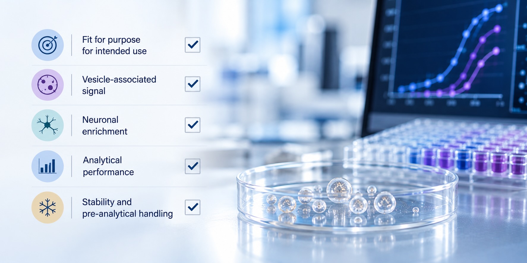

A translational team wants a neuronal vesicle readout in its first-in-human study. The assay runs well: repeat measurements of pooled plasma agree within 15%, and banked samples from patients separate from controls. Then a reviewer asks two questions. Tau and neurofilament circulate freely in plasma, so how do you know your signal comes from vesicles? And how do you know those vesicles came from neurons? Precision answers neither. That is where analytical validation for EV-based CNS assays has to do more work than a standard immunoassay validation.
Key takeaways
- EV-based CNS assays must answer the standard validation questions plus two more: is the signal vesicle-associated, and is it neuronal?
- Validation depth follows context of use. An exploratory signal in banked samples needs less than a pharmacodynamic readout or a trial enrichment marker.
- Neuronal vesicles are a minor fraction of plasma EVs, so the lower limit of quantification has to be set against baseline levels in the target population.
- Pre-analytical handling shifts absolute EV readouts between cohorts. Stability and handling data belong in the validation package.
What does analytical validation of an EV-based CNS assay require?
Analytical validation of an EV-based CNS assay requires fit-for-purpose evidence of precision, accuracy, parallelism, range and stability in plasma, plus two EV-specific proofs: that the signal comes from intact vesicles rather than free protein, and that those vesicles are enriched for neuronal origin.
The general definition hasn’t changed. Validation means presenting objective evidence that a method meets the requirements of its intended use. A practical guide written for neurology biomarker immunoassays lays out standard operating procedures for ten separate validation parameters, along with a reporting template [1]. The regulatory frame has moved, though. FDA’s guidance on bioanalytical method validation for biomarkers calls for a fit-for-purpose approach and points sponsors to the ICH M10 framework as a starting point, while expecting justification where M10 elements don’t fit a given biomarker platform [2].
EV-based CNS assays sit awkwardly inside that frame. An extracellular vesicle (EV) is a membrane-bound particle released by cells that carries surface proteins and internal cargo from its cell of origin; exosomes, the small vesicles formed in the endosomal pathway, are one subtype. A CNS assay built on EVs measures a protein on or inside a particle, from a neuronal subpopulation that makes up a small share of plasma vesicles, in a matrix that also contains the same protein in soluble form. Every standard parameter has to be reinterpreted with that in mind.
Start with context of use
Before any experiment, write down what decision the assay will support. The fit-for-purpose framework for drug development biomarkers treats validation as iterative: the evidence required grows as the data moves from exploratory research toward decisions with regulatory consequences [3].
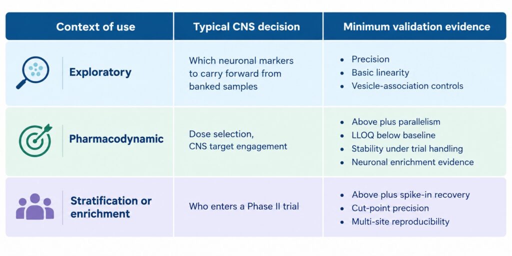
In CNS programs, a neuron-derived EV readout typically passes through three uses. It starts as an exploratory signal in banked samples. It becomes a pharmacodynamic marker in a first-in-human or Phase Ib study, where the question is whether the drug reached its target in the brain. In some programs it becomes a stratification or enrichment marker that decides who enters a Phase II trial. Teams that validate to the exploratory standard and then reuse the assay for a dose decision inherit every gap they skipped, usually at the point when a reviewer is least willing to wait.
Figure 1. Validation depth by context of use for EV-based CNS assays (illustrative).
The standard parameters, adapted for neuronal vesicles
Most items on the validation list carry over from conventional immunoassays. Each behaves differently when the analyte is a rare vesicle subpopulation.
Accuracy and recovery without a pure standard
There is no pure EV reference standard, and vesicles purified from cell culture don’t match the mixture found in plasma. Two kinds of spike-in material help. Recombinant EVs, engineered as trackable reference material with EV-like physical and biochemical properties, can be added to biofluids to measure recovery and to define variability introduced by sample handling between users [4]. For CNS assays, vesicles collected from human iPSC-derived neurons add a neuronal reference. In one neuronal EV enrichment method, spiked neuronal vesicles were recovered at 51% ± 10%, independent of input amount, and recovery varied by less than 13% across plasma from healthy donors and dementia patients [5]. Consistent recovery across disease and control plasma is the evidence that matters most, because a matrix effect that differs by diagnosis can manufacture a disease difference.
Parallelism and dilutional linearity in real plasma
Calibrators built in buffer rarely behave like vesicles in plasma. Parallelism testing, which compares the dilution curve of real samples with the calibration curve, is where matrix effects show up. Multiplex CNS panels need one more check: each analyte should read the same in the multiplex as it does on its own. In the published validation of one bead-based multiplex EV immunoassay, serially diluted plasma gave a linear signal, and simplex and multiplex formats showed similar linearity and detection range [6].
Range and lower limit of quantification
Neuron-derived vesicles make up only a minor fraction of circulating EVs [5]. Set the lower limit of quantification against the change the study needs to detect and against baseline levels in the target population, not against best-case sensitivity in buffer. A pharmacodynamic marker expected to move 20% after dosing is unusable if half the cohort sits below the quantifiable floor at baseline.
Precision
Precision for an EV-based CNS assay combines two sources of variance: the enrichment step and the measurement step. Report them together, because that is how the assay will run. In the neuronal enrichment method above, repeat processing of the same two plasma samples across five independent experiments gave coefficients of variation between 8.0% and 22.7% for p181-tau, total tau and amyloid-β42, and isolation was reproducible between operators and between labs [5]. We covered CV tiers and their sources in a separate post on building a reproducible plasma EV biomarker assay. The point here is narrower: precision is necessary, and it is the parameter EV-based CNS assays most often pass.
| Parameter | Conventional question | EV-based CNS question |
|---|---|---|
| Accuracy | Does the value match a known amount? | Are reference or iPSC-neuron vesicles recovered equally from patient and control plasma? |
| Selectivity | Do interferents change the result? | Does soluble tau, neurofilament or lipoprotein produce signal? |
| Specificity of origin | Not usually asked | Is the captured fraction enriched for neuronal material and depleted of blood-cell and liver material? |
| Parallelism | Do samples dilute like the calibrator? | Do vesicles in plasma dilute like the calibrator, in simplex and multiplex? |
| Range and LLOQ | What concentrations are quantifiable? | Is the neuronal subpopulation above LLOQ at baseline? |
| Precision | Is the result repeatable? | Is enrichment plus measurement repeatable across days, operators and labs? |
| Stability | Does the analyte survive storage? | Do vesicles and surface epitopes survive biobank freeze-thaw and trial storage? |
Proving the signal is vesicular and neuronal
Conventional guidance doesn’t name these two parameters, but reviewers of CNS programs increasingly ask about both.
Is it vesicular?
Many CNS analytes circulate in soluble form, and plasma lipoproteins overlap with small vesicles in size and density. An assay that captures soluble protein can show excellent precision and a plausible disease difference without measuring a single vesicle. MISEV2023, the International Society for Extracellular Vesicles’ minimal reporting standard, sets out the characterization expected for EV studies, including evidence that a measured signal is associated with vesicles [7]. In a validation package, that evidence usually comes from four controls:
- Detergent disruption. Lysing vesicle membranes should abolish or sharply reduce a signal that depends on intact particles.
- Vesicle depletion. Removing vesicles before the assay, by ultrafiltration or size-based separation, should remove the signal.
- Capture specificity. An isotype or non-specific capture control shows how much signal is background binding.
- Co-localization. Detecting the marker of interest together with a general vesicle marker, such as a tetraspanin, on the same captured particle shows both sit on one vesicle.
A design that builds co-localization into the readout makes these controls easier to pass. The LuminEV™ assay captures vesicles on antibody-coated beads and detects them with antibodies against surface tetraspanins, so a positive signal requires two surface proteins on the same particle. In its published validation, signal disappeared when vesicles were removed by ultrafiltration or disrupted with detergent, and the assay was validated for linearity, spike-in recovery, intra- and inter-assay precision and reproducibility between operators [6]. In plasma from 70 disease-free donors, the tetraspanins CD9, CD63 and CD81 accounted for 72%, 16% and 12% of the measured signal [6], a baseline that matters when tetraspanin detection is used to read neuronal subpopulations.
Is it neuronal?
Vesicle association is not enough for a CNS claim. The assay also has to show that the captured fraction is enriched for neuronal material. The practical test compares cell-type signatures before and after enrichment. In the neuronal enrichment validation cited above, erythrocyte, platelet and liver mRNA in the enriched fraction fell to under 6% of plasma levels while neuronal mRNA was retained, and neuron-specific proteins were enriched compared with an isotype capture control [5]. Both studies cited in this section were led by NeuroDex scientists with academic collaborators, so treat them as the developer’s own evidence and ask for the same controls from any platform you evaluate.
Stability and pre-analytics are validation parameters, too
CNS biomarker programs lean heavily on biobanks and multi-site trials, which makes pre-analytical variance a validation problem rather than a logistics one. The ISEV Blood EV Task Force counted hundreds of pre-analytical protocols and more than forty variables in use, and responded with MIBlood-EV, a reporting tool covering blood collection, processing, storage and plasma quality [8].
The effect shows up in real CNS data. In the multi-cohort neuronal EV study above, differences between Alzheimer’s patients and controls held their direction across cohorts, but absolute levels of amyloid-β42 and proBDNF varied widely between cohorts, which the authors attributed partly to cohort-specific pre-analytical conditions [5]. For a pharmacodynamic marker read against a fixed threshold, that kind of shift is the difference between a positive and a negative result.
Storage adds its own drift. In a two-year comparison of storage buffers and temperatures, EVs kept in phosphate-buffered saline showed drastically reduced recovery at every temperature tested, starting within days; buffers supplemented with albumin and trehalose largely prevented the loss [9]. A systematic study of storage effects on isolated EVs and platelet-free plasma found that none of eight conditions tested prevented storage-related changes, with evidence of vesicle fusion [10].
For EV-based CNS assays, that translates into four experiments:
- Freeze-thaw cycles on whole plasma, at the number of cycles a biobank sample is likely to have seen.
- Bench-top time between thaw and assay, matched to real plate-handling times.
- Long-term storage at the temperature the trial will use, with timepoints spanning the study duration.
- Processed-sample stability if enriched vesicles, diluted samples or captured beads wait before reading.
Measure stability with the assay itself. Particle counts can stay flat while the surface epitope an assay captures degrades, and only the assay’s own readout will show that.
The checklist, and what to ask of any EV platform
The table collects the parameters into one checklist for EV-based CNS assays. Use it to build an in-house validation plan, or to evaluate EV biomarker assay services before committing biopharma samples to them.
| Parameter | Test | Evidence to see | Needed for |
|---|---|---|---|
| Context of use | Written statement of decision supported | Defined marker category and acceptable error | All |
| Vesicle association | Detergent disruption, depletion, isotype control, co-localization | Signal lost when vesicles are removed or lysed | All |
| Neuronal origin | Cell-type signatures before vs. after enrichment | Neuronal markers retained; blood-cell and liver markers depleted | All |
| Precision | Intra-, inter-assay, inter-operator and inter-lab runs including enrichment | CVs within pre-set limits for the context of use | All |
| Dilutional linearity | Serial dilution of real plasma | Linear response across working range | All |
| Parallelism | Sample vs. calibrator curves; simplex vs. multiplex | Parallel curves; matching simplex and multiplex values | Pharmacodynamic, stratification |
| Range and LLOQ | LLOQ against baseline levels in the target population | Most baseline samples above LLOQ | Pharmacodynamic, stratification |
| Accuracy and recovery | Spike-in of reference or iPSC-neuron vesicles into patient and control plasma | Consistent recovery regardless of diagnosis | Stratification |
| Stability | Freeze-thaw, bench-top, long-term, processed sample | Signal retained under biobank and trial handling | Pharmacodynamic, stratification |
| Pre-analytical reporting | MIBlood-EV-style record of collection and processing | Documented, reproducible handling at every site | All |
| Multi-site reproducibility | Shared samples run across labs or operators | Agreement within pre-set limits | Stratification |
When the assay comes from a vendor, the same table becomes a list of questions. Has the platform shown that soluble tau or neurofilament doesn’t produce signal? Does it publish neuronal enrichment data, not just vesicle characterization? Is parallelism shown in plasma rather than buffer? Were simplex and multiplex formats compared? Is stability data available for the storage conditions your trial uses? A platform that answers with published data saves months of in-house work. For teams running these checks in their own lab, the LuminEV™ Research Kit includes the multiplex format and vesicle-association controls described above.
The field’s next step is shared reference material. Recombinant vesicles, iPSC-neuron vesicles and blood-specific reporting standards already exist. What’s missing is agreement on which reference vesicles, at what concentrations, every EV-based CNS assay should be tested against. Once that exists, comparing neuronal EV results between labs and between trials will stop depending on trust. Until then, each assay team carries the burden of showing that what it measures is a vesicle and that the vesicle came from a neuron. That proof is also the foundation of any bridging study from animal model to first-in-human that relies on a blood-based CNS biomarker.
References
- Andreasson U, Perret-Liaudet A, van Waalwijk van Doorn LJC, et al. A practical guide to immunoassay method validation. Front Neurol. 2015;6:179. https://doi.org/10.3389/fneur.2015.00179
- U.S. Food and Drug Administration. Bioanalytical Method Validation for Biomarkers: Guidance for Industry. 2025. https://www.fda.gov/regulatory-information/search-fda-guidance-documents/bioanalytical-method-validation-biomarkers
- Lee JW, Devanarayan V, Barrett YC, et al. Fit-for-purpose method development and validation for successful biomarker measurement. Pharm Res. 2006;23(2):312-328. https://doi.org/10.1007/s11095-005-9045-3
- Geeurickx E, Tulkens J, Dhondt B, et al. The generation and use of recombinant extracellular vesicles as biological reference material. Nat Commun. 2019;10(1):3288. https://doi.org/10.1038/s41467-019-11182-0
- Eitan E, Thornton-Wells T, Elgart K, et al. Synaptic proteins in neuron-derived extracellular vesicles as biomarkers for Alzheimer’s disease: novel methodology and clinical proof of concept. Extracell Vesicles Circ Nucleic Acids. 2023;4:133-150. https://doi.org/10.20517/evcna.2023.13
- Tordoff E, Allen J, Elgart K, et al. A novel multiplexed immunoassay for surface-exposed proteins in plasma extracellular vesicles. J Extracell Vesicles. 2024;13(11):e70007. https://doi.org/10.1002/jev2.70007
- Welsh JA, Goberdhan DCI, O’Driscoll L, et al. Minimal information for studies of extracellular vesicles (MISEV2023): from basic to advanced approaches. J Extracell Vesicles. 2024;13(2):e12404. https://doi.org/10.1002/jev2.12404
- Lucien F, Gustafson D, Lenassi M, et al. MIBlood-EV: minimal information to enhance the quality and reproducibility of blood extracellular vesicle research. J Extracell Vesicles. 2023;12(12):e12385. https://doi.org/10.1002/jev2.12385
- Görgens A, Corso G, Hagey DW, et al. Identification of storage conditions stabilizing extracellular vesicles preparations. J Extracell Vesicles. 2022;11(6):e12238. https://doi.org/10.1002/jev2.12238
- Gelibter S, Marostica G, Mandelli A, et al. The impact of storage on extracellular vesicles: a systematic study. J Extracell Vesicles. 2022;11:e12162. https://doi.org/10.1002/jev2.12162