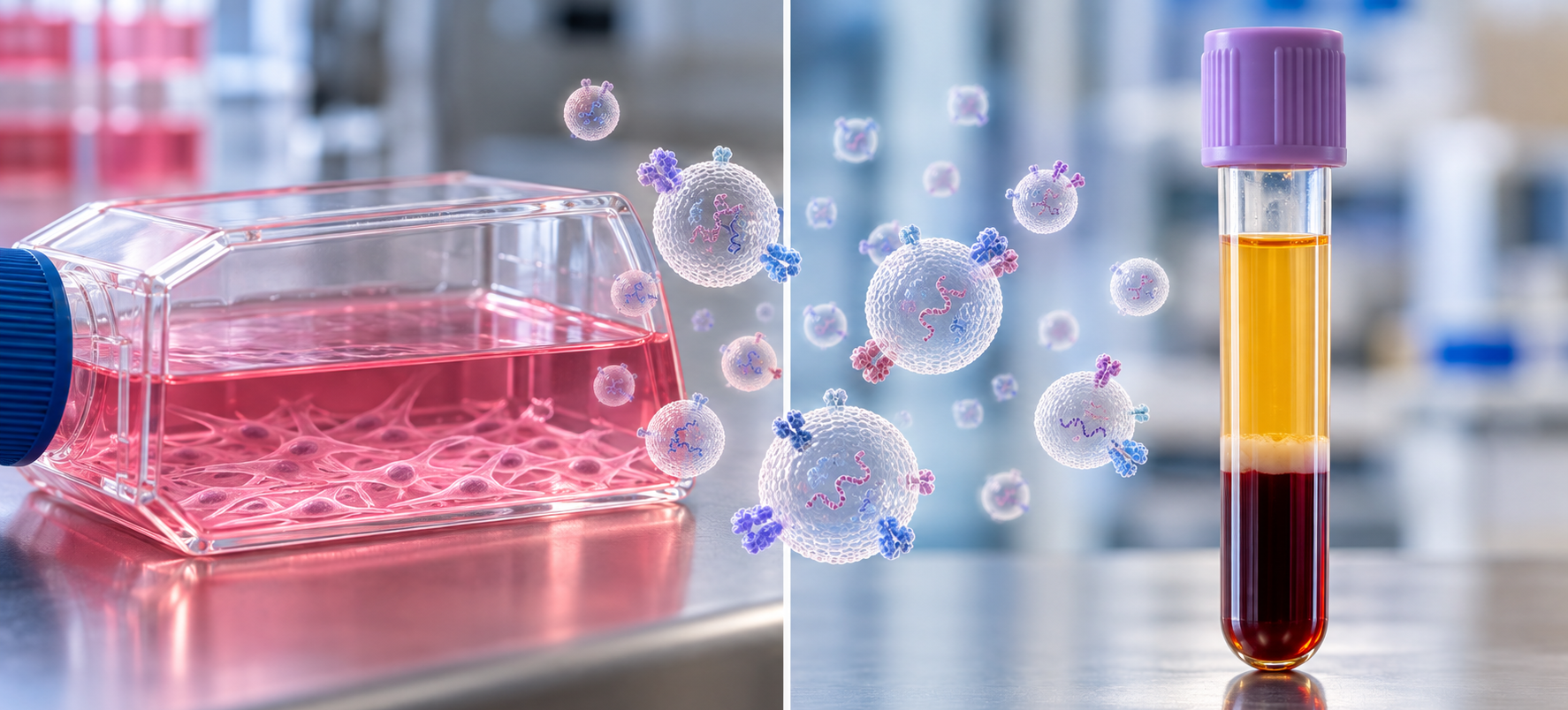

A lab validates an extracellular vesicle (EV) surface marker in conditioned media from cultured neurons. The signal is clean, dose-responsive, and reproducible across passages. Then the same assay goes into patient plasma, and the signal nearly disappears into background. The biology did not change. The matrix did. Most EV methods are developed in culture and then carried into blood, but the two starting materials differ in almost every variable that decides what an assay measures.
Key takeaways
- Conditioned media holds EVs from one cell type plus serum-derived contaminants; plasma holds EVs from many tissues inside a matrix dominated by lipoproteins and soluble proteins.
- Even commercially EV-depleted FBS can leave hundreds of millions of bovine EVs per mL in culture medium.
- Culture conditions such as oxygen level change EV release, while blood collection and processing introduce dozens of pre-analytical variables.
- Markers defined in culture need to be re-validated in plasma, ideally with methods that limit isolation-driven bias.
How are plasma EVs different from cell culture EVs?
Plasma EVs and cell culture EVs differ in three ways: plasma contains vesicles from many cell types rather than one, sits in a matrix crowded with lipoproteins and proteins rather than defined medium, and carries pre-analytical variability from blood collection that culture experiments largely control.
Cell culture is how most researchers first meet EVs. An international survey of ISEV members found that 83% of respondents used conditioned culture media as their starting material [4]. That makes sense: culture offers a known cell of origin, controllable conditions, and enough volume to isolate, characterize, and repeat.
Plasma offers none of those conveniences. What it offers instead is clinical relevance. If a vesicle marker is ever going to report on disease in people, it has to work in blood. The problem is that a method tuned for one matrix can fail quietly in the other, and the failure often looks like a biological negative result rather than a technical one.
Figure 1 summarizes the main differences. The sections below take each in turn.
| Variable | Conditioned culture media | Blood plasma |
|---|---|---|
| Cellular sources | One cell type (or a defined co-culture) | Many tissues and blood cells at once |
| Main non-target particles | Bovine EVs, RNA, and aggregates from FBS | Lipoproteins and abundant soluble proteins |
| Biggest controllable variables | Serum type, confluence, oxygen, collection time | Tube type, time to processing, platelet removal, storage |
| Typical sample volume | Tens of mL or more | Often 1 mL or less per aliquot |
| Reporting framework | MISEV2023 | MISEV2023 plus MIBlood-EV |
What’s actually in a plasma EV preparation
Lipoproteins outnumber vesicles
Plasma is not a dilute EV suspension. Circulating lipoproteins are estimated to be around six orders of magnitude more abundant than circulating EVs, and they overlap with EVs in size and density [2]. That overlap means size- and density-based isolation methods tend to co-isolate them. It also means lipid dyes and particle counters cannot reliably tell the two apart. Researchers have also reported evidence of EVs binding and fusing with lipoprotein-like structures in plasma [2], which complicates the picture further.
For a researcher coming from culture, the practical consequence is simple. A particle count that was meaningful in conditioned media may be mostly lipoprotein in plasma. Protein-level readouts tied to EV-specific surface markers hold up better than counts or lipid labels.
Many cellular sources, one tube
Plasma EVs come from blood cells, endothelium, and solid tissues together. When researchers profiled plasma from 70 disease-free donors with a multiplexed surface protein immunoassay, CD9 accounted for 72% of the tetraspanin signal, CD63 for 16%, and CD81 for 12% [1]. The same assay detected vesicles carrying markers of erythrocytes, neurons, and macrophages [1].
That mixture has a direct consequence for anyone moving a marker out of culture. A tetraspanin profile measured on EVs from one cultured cell type tells you little about what the same tetraspanins will look like in plasma, where the signal is a blend of many sources. A tissue-specific signal that dominated conditioned media may be a small fraction of the plasma EV pool. For more on the three canonical tetraspanins and what they do and do not tell you, see our tetraspanin guide.
What’s actually in a conditioned-media preparation
The serum problem
Cultured cells usually need serum, and fetal bovine serum (FBS) is full of its own EVs. In one quantitative test, culture medium with 10% untreated FBS contained about 2.6 × 10¹⁰EVs per mL before a single cell was added [3]. Depletion helps less than many researchers assume. An 18-hour ultracentrifugation and a commercial EV-depleted FBS were both tested. The commercial product removed the most, about 75% of FBS EVs, yet still left roughly 6.9 × 10⁸ EVs per mL in the medium. Both depletion approaches also reduced the growth and viability of primary astrocytes [3].
Bovine EVs are not the only contaminant. FBS also carries RNA and protein aggregates that can co-isolate with cell-derived vesicles and alter their apparent cargo [5]. Because EV-depleted FBS is usually prepared by ultracentrifugation or precipitation, it can also differ from standard serum in lipoprotein content. That matters because lipoprotein levels in culture medium change the effects EVs have on recipient cells [2].
Culture conditions change what cells release
Culture gives control, but every controlled variable shifts EV output. In breast cancer cell lines, moderate (1% O2) and severe (0.1% O2) hypoxia increased exosome release, an effect that depended on HIF-1α signaling [6]. More broadly, culture conditions themselves, starting with whether and how serum is used, shape what cells release and what ends up in the EV preparation [5]. A marker that looks stable under one set of conditions may simply reflect them.
Pre-analytics in each matrix
In culture, the variables are mostly upstream and under the experimenter’s control. In blood, many are set before the sample reaches the lab. The ISEV Blood EV Task Force counted hundreds of pre-analytical protocols in use and more than forty variables that can affect blood EV results. It responded with MIBlood-EV, a reporting tool that records how plasma and serum were collected and prepared and how their quality was assessed [7]. Key variables include collection tube, time to processing, centrifugation steps that remove platelets, and freeze-thaw history.
Storage affects both matrices. In a systematic study, −80°C storage reduced EV concentration and purity over time, and no storage additive tested prevented it. EV size remained stable when the starting fluid was stored but increased when isolated EVs were stored [9]. The lesson holds for plasma and conditioned media alike: freeze the fluid, not the isolate, and record how long it sat.
What to record, matrix by matrix
Recording these variables costs little at collection time and is nearly impossible to reconstruct later. For conditioned media, record:
- serum type, concentration, and depletion method (or serum-free formulation);
- cell passage number and confluence at the start of conditioning;
- conditioning time and oxygen level;
- how medium was cleared of cells and debris before storage.
For plasma, record:
- collection tube and anticoagulant;
- time from draw to first centrifugation;
- centrifugation protocol, including any second spin to remove platelets;
- hemolysis or lipemia assessment;
- freeze date, storage temperature, and freeze-thaw count.
For both matrices, MISEV2023 sets the baseline for what to report, from source and collection to separation and characterization [8]. For blood, MIBlood-EV adds the pre-analytical detail that MISEV alone does not capture.
Why isolation choices widen the gap
Isolation methods behave differently in each matrix. A protocol that gives clean pellets from conditioned media can co-isolate large amounts of lipoprotein from plasma, because the contaminants are different. Methods tuned to remove serum-derived bovine EVs do nothing for lipoproteins, and methods tuned to remove lipoproteins may lose the small tissue-specific populations that made plasma interesting in the first place. Every added isolation step is another point where recovery can differ between samples, sites, and matrices. Our overview of EV detection and analysis methods covers the trade-offs of each approach in more detail.
One way to narrow the gap is to measure surface proteins directly in the starting fluid, without a separate isolation step. Sandwich immunoassays that require two distinct surface proteins on the same particle report on intact vesicles while ignoring free proteins and most lipoproteins. The LuminEV assay was developed on this principle and validated in purified EVs, cell culture media, and blood plasma [1]. Because the same assay runs in both matrices without a matrix-specific isolation step, it removes one source of variation when a marker moves from culture to plasma. It does not remove the biological differences, which still need to be designed around.
Designing studies that bridge both
Culture and plasma answer different questions. Culture is best for defining what a cell type releases and how that changes with a stimulus. Plasma is where you test whether that release is detectable, specific, and informative in people. A bridging study uses each for what it does well.
Define the marker in culture, then test it where it will be used
Use culture to establish that a surface marker is present on vesicles from the cell type of interest. Do the culture work in serum-free medium where the cells tolerate it, or include a medium-only control processed identically, so bovine signal can be subtracted. Then move to plasma early, with small pilot sets, before investing in larger culture experiments that may not translate. For how pilot data should feed into larger plasma studies, see Building a Reproducible Plasma Biomarker Assay.
Use matrix-matched controls
In plasma, include EV-depleted plasma or detergent-treated samples to confirm that the signal is vesicle-associated. Include samples spanning the expected range of lipid levels if lipoproteins could interfere. In culture, include unconditioned medium. In both, run a positive control vesicle preparation across plates and batches.
Plan for the volume you will actually have
Culture experiments can generate tens of milliliters of conditioned medium per condition. Plasma studies rarely have that luxury: banked aliquots are often 1 mL or less, and every analyte competes for the same tube. Methods that need a large input for isolation before measurement may be fine in culture and impractical in a cohort. Plan the plasma assay around the volume available per participant, and favor multiplexed readouts that measure several markers from one aliquot. We showed what that looks like for neurodegeneration markers in Tau, p-tau181, TDP-43, and α-Synuclein From 1 mL of Plasma.
Normalize thoughtfully
Normalizing a tissue-specific marker to total EV signal works differently in each matrix. In conditioned media, total tetraspanin signal mostly reflects one cell type’s output. In plasma, it reflects the whole circulating pool, dominated by blood cells. Reporting both the raw and the normalized values lets readers judge whether a change comes from the marker or the denominator.
| Usually transfers | Usually does not transfer |
|---|---|
| Whether a protein is present on vesicles from a given cell type | The absolute level of that protein in a plasma EV population |
| Direction of change in response to a well-defined stimulus | Effect size, once the signal is diluted by other cellular sources |
| Antibody pair performance on purified vesicles | Specificity in the presence of lipoproteins and soluble proteins |
| Basic assay precision on a reference preparation | Pre-analytical stability, which must be tested in the target matrix |
Plasma is often described as conditioned media that is simply messier. That framing undersells it. Plasma holds vesicles from every tissue at once, including small populations from organs that cannot be sampled directly. Recent work on enriching tissue-specific EVs from circulation treats that mixture as the opportunity rather than the obstacle [10], a theme we take up in The Next Decade of EV Liquid Biopsy Will Be Decided by Tissue of Origin. The methods that make plasma EV research reproducible will be the ones built for that mixture from the start, not adapted to it after the culture experiments are done.
References
- Tordoff E, Allen J, Elgart K, et al. A novel multiplexed immunoassay for surface-exposed proteins in plasma extracellular vesicles. J Extracell Vesicles. 2024;13(11):e70007. https://doi.org/10.1002/jev2.70007
- Busatto S, Yang Y, Iannotta D, Davidovich I, Talmon Y, Wolfram J. Considerations for extracellular vesicle and lipoprotein interactions in cell culture assays. J Extracell Vesicles. 2022;11(4):e12202. https://doi.org/10.1002/jev2.12202
- Lehrich BM, Liang Y, Khosravi P, Federoff HJ, Fiandaca MS. Fetal bovine serum-derived extracellular vesicles persist within vesicle-depleted culture media. Int J Mol Sci. 2018;19(11):3538. https://doi.org/10.3390/ijms19113538
- Lehrich BM, Liang Y, Fiandaca MS. Foetal bovine serum influence on in vitro extracellular vesicle analyses. J Extracell Vesicles. 2021;10:e12061. https://doi.org/10.1002/jev2.12061
- Urzì O, Olofsson Bagge R, Crescitelli R. The dark side of foetal bovine serum in extracellular vesicle studies. J Extracell Vesicles. 2022;11:e12271. https://doi.org/10.1002/jev2.12271
- King HW, Michael MZ, Gleadle JM. Hypoxic enhancement of exosome release by breast cancer cells. BMC Cancer. 2012;12:421. https://doi.org/10.1186/1471-2407-12-421
- Lucien F, Gustafson D, Lenassi M, et al. MIBlood-EV: minimal information to enhance the quality and reproducibility of blood extracellular vesicle research. J Extracell Vesicles. 2023;12(12):e12385. https://doi.org/10.1002/jev2.12385
- Welsh JA, Goberdhan DCI, O’Driscoll L, et al. Minimal information for studies of extracellular vesicles (MISEV2023): from basic to advanced approaches. J Extracell Vesicles. 2024;13(2):e12404. https://doi.org/10.1002/jev2.12404
- Gelibter S, Marostica G, Mandelli A, et al. The impact of storage on extracellular vesicles: a systematic study. J Extracell Vesicles. 2022;11(2):e12162. https://doi.org/10.1002/jev2.12162
- Pierri B, Eitan E, Witwer KW, Re DB, Baccarelli AA, Wu H. Tissue-specific extracellular vesicles enriched from circulation: exploring the liquid biopsy perspective. J Extracell Biol. 2026;5(2):e70106. https://doi.org/10.1002/jex2.70106