
Introduction: The Silent Messengers of the Cell
In the intricate symphony of cellular life, communication is paramount. For decades, scientific understanding centered on direct cell-to-cell contact or the action of secreted soluble molecules. However, a groundbreaking revelation has reshaped our view: cells are also capable of releasing nanoscale vesicles, microscopic envelopes brimming with vital information. These are extracellular vesicles (EVs), the silent messengers that traverse bodily fluids, influencing a vast array of biological processes. Far from being mere cellular debris, EVs are sophisticated carriers, orchestrating complex interactions that impact health and disease. This article delves into the fascinating world of extracellular vesicles, exploring their formation, their diverse cargoes, their roles in physiological and pathological states, and their burgeoning potential to revolutionize medicine.
The Silent Messengers of the Cell
The discovery and subsequent exploration of extracellular vesicles have opened a new frontier in biological research. These minute particles, present in virtually all biological fluids including blood, urine, saliva, and cerebrospinal fluid, serve as critical conduits for intercellular communication. They are not passive byproducts of cellular activity but actively secreted entities, meticulously packaged with molecular information from their parent cell. Their presence in these fluids suggests a widespread and constant dialogue between cells throughout the body. Understanding this dialogue is key to deciphering numerous biological phenomena, from immune responses to tissue regeneration and the progression of complex diseases.
What Are Extracellular Vesicles (EVs)?
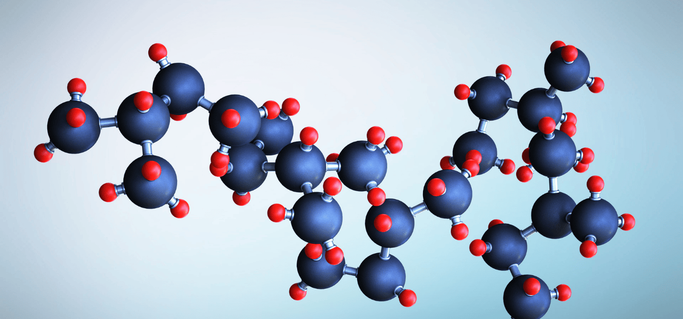
![]() Anatomy of an Extracellular Vesicle: A cross-section revealing the lipid bilayer membrane and the diverse molecular cargo it transports, including proteins and nucleic acids.
Anatomy of an Extracellular Vesicle: A cross-section revealing the lipid bilayer membrane and the diverse molecular cargo it transports, including proteins and nucleic acids.
Extracellular vesicles (EVs) are a heterogeneous population of lipid bilayer-enclosed particles released by cells into their extracellular environment. They range in size from approximately 30 nanometers to over a micrometer and are found in nearly all body fluids and secreted by virtually all cell types. At their core, EVs are designed to carry and deliver biological information. This information can include a diverse array of molecules such as proteins, lipids, messenger RNAs (mRNAs), microRNAs (miRNAs), and even DNA fragments. Their primary function is to facilitate intercellular communication, allowing cells to influence the behavior and function of recipient cells without direct physical contact. This communication can occur locally, between adjacent cells, or systemically, as EVs travel through the bloodstream or other bodily fluids to distant tissues.
The Ubiquitous Role in Intercellular Communication
Intercellular communication is the bedrock of multicellular life, enabling coordinated tissue function, development, and adaptation. Extracellular vesicles have emerged as a pivotal mechanism for this communication. They act as sophisticated couriers, shuttling molecular messages that can alter the gene expression, protein synthesis, and overall phenotype of recipient cells. This intercellular dialogue is fundamental to maintaining physiological homeostasis, responding to environmental cues, and orchestrating complex biological processes such as immune surveillance, blood vessel formation, and nervous system function. The ability of cells to secrete and receive information via EVs provides a nuanced layer of biological regulation, allowing for precise control and sophisticated responses within the organism.
Why Understanding EV Biogenesis Matters for Therapy
The processes by which cells generate and release extracellular vesicles, known as EV biogenesis, are far from random. They are intricate, regulated pathways that determine the size, composition, and ultimately, the function of the released EVs. Understanding these biogenesis pathways is not merely an academic pursuit; it is fundamentally important for harnessing EVs for therapeutic purposes. By comprehending how specific cargoes are sorted into EVs and how different types of EVs are formed, scientists can begin to engineer these vesicles for targeted drug delivery, regenerative medicine, and immunomodulation. Manipulating EV biogenesis allows for the production of therapeutic EVs with enhanced efficacy, specificity, and safety profiles, paving the way for next-generation medical interventions.
Article Overview: From Formation to Future Medicine
This article aims to provide a comprehensive yet accessible exploration of extracellular vesicles. We will begin by dissecting the diverse types of EVs and the intricate mechanisms of their biogenesis. Subsequently, we will delve into the molecular cargoes that equip these vesicles for their signaling roles and examine their widespread functions in both health and disease, with a particular focus on their implications in cancer. Finally, we will explore the revolutionary therapeutic potential of EVs, from their use as natural drug delivery systems to their applications in regenerative medicine and as powerful diagnostic biomarkers, while also addressing the challenges that lie ahead in translating this promise into clinical reality.
The Diverse World of Extracellular Vesicles: A Classification
While collectively termed extracellular vesicles, this population is not monolithic. It comprises distinct subtypes, each characterized by its biogenesis pathway, size, and typical cargo. Understanding these differences is crucial for appreciating their varied roles and therapeutic applications.
Exosomes: The Endosomal Originators
Exosomes are a well-studied class of EVs, typically ranging from 30 to 150 nanometers in diameter. Their defining characteristic is their origin from the endosomal pathway. Specifically, they are formed as intraluminal vesicles (ILVs) within multivesicular bodies (MVBs), which are late endosomal compartments. These MVBs then fuse with the plasma membrane, releasing their contained ILVs into the extracellular space as exosomes. This endosomal origin results in a unique molecular signature, often enriched with proteins involved in endocytosis, MVB formation, and membrane trafficking, such as tetraspanins (CD9, CD63, CD81) and Alix. The cargo within exosomes is carefully sorted during their formation, reflecting the specific cellular state and needs of the parent cell.
Microvesicles (Ectosomes): Direct Budding from the Plasma Membrane
Microvesicles, also known as ectosomes, represent another major class of EVs. Unlike exosomes, microvesicles are generated through direct outward budding of the cell’s plasma membrane. This process involves the outward protrusion and subsequent fission of the plasma membrane, releasing vesicles that are generally larger than exosomes, typically ranging from 100 to 1000 nanometers in diameter. The molecular composition of microvesicles reflects their plasma membrane origin, often carrying surface proteins such as adhesion molecules, cytoskeletal proteins like actin and myosin, and components of the signaling machinery found at the cell surface. Their formation is often linked to cellular activation, stress, or apoptosis, and their cargo is highly dependent on the state of the plasma membrane at the time of budding.
Other EV Types: Apoptotic Bodies and Beyond
Beyond exosomes and microvesicles, other EV-like structures exist, with apoptotic bodies being a notable example. Apoptotic bodies are larger vesicles, often exceeding one micrometer, that are released by cells undergoing programmed cell death (apoptosis). They contain cellular contents, including nuclear DNA, fragmented organelles, and cytoplasmic components, and play a role in clearing cellular debris and modulating the immune response during apoptosis. While historically distinguished, the lines between these categories can blur, and the classification of EVs remains an active area of research, with emerging evidence suggesting further heterogeneity and complex origins.
Key Distinctions: Size, Biogenesis, and Composition
The primary distinctions between EV subtypes lie in their origin, size, and molecular composition. Exosomes arise from intracellular endosomal compartments and are released via MVB fusion with the plasma membrane, typically exhibiting smaller sizes. Microvesicles, in contrast, are shed directly from the plasma membrane through budding, generally resulting in larger particles. These distinct biogenesis pathways lead to differential enrichment of specific molecules. Exosomes often carry endosomal-associated proteins and miRNAs involved in gene regulation, while microvesicles can bear surface receptors and cytoplasmic proteins reflective of their plasma membrane origin. This cargo specificity dictates their distinct functional roles and therapeutic applicability.
EV Biogenesis: Unraveling How Cells Create and Release Their Cargo
The creation and release of extracellular vesicles are sophisticated cellular processes that are tightly regulated. Understanding the mechanisms of EV biogenesis is critical for controlling their production and tailoring their therapeutic potential.
Exosome Biogenesis: The Intricate Endosomal Pathway
Exosome biogenesis begins with the inward budding of the endosomal membrane, forming small vesicles within the endosome itself. These vesicles mature into multivesicular bodies (MVBs). The cargo destined for exosomes, including specific proteins and nucleic acid molecules, is sorted and packaged into these intraluminal vesicles (ILVs) during their formation. This sorting process is complex and involves various cellular mechanisms, such as the endosomal sorting complex required for transport (ESCRT) machinery, ubiquitination signals, and specific lipid or protein interactions. Once formed, MVBs can either fuse with lysosomes for degradation or traffic to the plasma membrane to fuse and release their ILVs as exosomes into the extracellular space.
Microvesicle (Ectosome) Biogenesis: Direct Membrane Shedding
The biogenesis of microvesicles, or ectosomes, is a more direct process originating from the cell’s outer boundary. It involves the outward budding of the plasma membrane, driven by changes in membrane fluidity and the coordinated action of cytoskeletal elements, particularly actin. As the membrane protrudes, it encapsulates cytoplasmic components, including proteins, lipids, and RNA. The vesicle then pinches off from the parent cell, forming a microvesicle. The specific cargo within microvesicles is influenced by the state of the plasma membrane and the underlying cytoplasm at the moment of budding, making them sensitive indicators of cellular physiological or pathological conditions.
The Regulation of EV Biogenesis: A Coordinated Process
The biogenesis of both exosomes and microvesicles is not a passive process but is actively regulated by cellular signaling pathways, metabolic status, and external stimuli. Factors such as nutrient availability, stress, hypoxia, and inflammatory signals can all influence the rate of EV production and the composition of their cargo. This regulatory control allows cells to fine-tune their communication networks in response to their environment. The ability to influence these regulatory pathways offers a promising avenue for enhancing the therapeutic benefits of EVs, for example, by stimulating cells to produce EVs with a specific therapeutic content.
The Message Within: Decoding EV Cargo and Its Functional Significance
The true power of extracellular vesicles lies in the molecular information they carry. This diverse content, or cargo, dictates their function and their impact on recipient cells.
A Multitude of Cargoes: The Molecular Blueprint
Extracellular vesicles are packed with a remarkable variety of biomolecules, forming a complex molecular blueprint. This cargo includes a vast repertoire of proteins, ranging from enzymes and signaling molecules to structural components. They also carry nucleic acid payloads, such as mRNAs that can be translated into new proteins in recipient cells, and miRNAs that can regulate gene expression post-transcriptionally. Lipids, carbohydrates, and even DNA fragments can also be found within EVs. The specific composition of this cargo is highly cell-type specific and varies depending on the physiological or pathological state of the parent cell, providing a snapshot of cellular activity.
Cargo Sorting Mechanisms: How Specificity is Achieved
Cells employ sophisticated mechanisms to ensure that specific molecules are sorted into the correct type of extracellular vesicle. For exosomes, this sorting process often involves the ESCRT pathway, ubiquitination tags on proteins, and the specific enrichment of certain lipid species within the forming ILVs. Microvesicle cargo is more directly representative of the cell surface and cytoplasm at the time of budding, influenced by membrane domains and cytoskeletal interactions. This selective packaging ensures that the EVs deliver relevant and functional information to recipient cells, enabling precise and targeted intercellular communication.
Intercellular Communication: How EVs Deliver Their Message
Once released, extracellular vesicles can interact with recipient cells through several mechanisms. They can bind to receptors on the surface of target cells, triggering intracellular signaling cascades. They may also be internalized by recipient cells through endocytosis or direct fusion with the plasma membrane, delivering their entire molecular cargo for processing within the cell. This ability to transfer functional molecules allows EVs to reprogram recipient cells, influencing processes such as gene expression, protein synthesis, and cellular differentiation. This mode of intercellular communication is fundamental to tissue development, immune responses, and the modulation of disease states.
EV Functions in Health and Disease: More Than Just Waste Disposal
Extracellular vesicles are not mere cellular byproducts but active participants in maintaining health and driving disease progression. Their roles are diverse and profound.
Physiological Roles: Maintaining Homeostasis
In healthy organisms, EVs play critical roles in maintaining homeostasis. They are involved in immune system regulation, facilitating communication between immune cells and modulating inflammatory responses. They contribute to tissue repair and regeneration by delivering growth factors and signaling molecules that promote cell proliferation and differentiation. EVs also play a part in cardiovascular health, neuroprotection, and metabolic regulation. This constant exchange of information via EVs helps to coordinate complex physiological processes and ensure the smooth functioning of the organism.
Pathological Roles: Contributing to Disease Progression
Dysregulated EV production and function are implicated in the pathogenesis of numerous diseases. In cancer, EVs can promote tumor growth, invasion, and metastasis by delivering factors that stimulate angiogenesis, suppress anti-tumor immunity, and prepare distant sites for colonization. They can also mediate drug resistance. In neurodegenerative diseases like Alzheimer’s and Parkinson’s, EVs can contribute to the spread of misfolded proteins and neuroinflammation. EVs also play significant roles in cardiovascular diseases, autoimmune disorders, and infectious diseases, highlighting their dual nature as facilitators of both health and pathology. Understanding these pathological roles is crucial for developing targeted therapeutic interventions.
Unlocking Therapeutic Potential: EVs as Next-Generation Medicines
The remarkable properties of extracellular vesicles position them as a promising new class of therapeutics and diagnostic tools. Their ability to carry and deliver specific molecular cargoes, their inherent biocompatibility, and their natural signaling capabilities open up vast possibilities.
EVs as Natural Drug Delivery Vehicles
The inherent structure of EVs makes them ideal candidates for drug delivery. They are naturally occurring nanoparticles, biocompatible, and possess low immunogenicity compared to synthetic carriers. Scientists can engineer EVs to carry therapeutic proteins, nucleic acids, or small molecule drugs. Furthermore, EVs can be modified to target specific cell types or tissues, enhancing drug efficacy and reducing off-target effects. For instance, EVs can be loaded with chemotherapy drugs to target cancer cells, or with gene-editing tools to correct genetic defects. Their ability to cross biological barriers, such as the blood-brain barrier, further enhances their potential for delivering therapeutics to previously inaccessible sites. The ongoing research in this area aims to provide robust methods to support these applications.
Regenerative Medicine: Harnessing EV Repair Capabilities
Extracellular vesicles, particularly those derived from mesenchymal stem cells (MSCs), have demonstrated significant potential in regenerative medicine. These EVs possess inherent regenerative properties, delivering bioactive molecules that can stimulate tissue repair, promote cell survival, and reduce inflammation. They can be utilized in applications aimed at repairing damaged heart tissue after a heart attack, regenerating cartilage in osteoarthritis, or promoting wound healing. The global regenerative medicine market was valued at USD 40.51 billion in 2024 and is expected to reach USD 236.28 billion by 2032, growing at a CAGR of 24.66% from 2025-2032 SNS Insider, underscoring the vast economic and therapeutic landscape for EV-based regenerative therapies.
Immunomodulation and Cancer Therapy
Extracellular vesicles are increasingly being explored for their roles in immunomodulation and cancer therapy. By engineering EVs, researchers can design them to deliver immunomodulatory payloads that either stimulate an anti-tumor immune response or dampen excessive inflammation. In cancer treatment, the oncology segment held the major market share of 40% in exosome therapeutics by therapeutic application in 2024 Precedence Research. This statistic highlights the significant focus on leveraging EVs to combat malignancies. EVs can be loaded with mRNA vaccines to prime the immune system against cancer cells, or engineered to deliver chemotherapeutic agents directly to tumors, minimizing systemic toxicity.
EVs as Biomarkers: Early Detection and Disease Monitoring
The presence and composition of EVs in body fluids can serve as invaluable biomarkers for disease detection, diagnosis, and monitoring. The EV-based liquid biopsy market is valued at USD 102.13 million in 2024 and is projected to reach USD 572.43 million by 2034, growing at a 19.0% CAGR Exactitude Consultancy. This statistic underscores the burgeoning utility of EVs in non-invasive diagnostics. By analyzing the EVs found in blood, urine, or other samples, clinicians can detect early signs of diseases like cancer, neurodegenerative disorders, and cardiovascular conditions. Changes in EV content and concentration can also track disease progression, response to treatment, and predict patient outcomes, providing crucial diagnostic information.
Translating EVs to the Clinic: Challenges and the Road Ahead
Despite the immense promise, translating the therapeutic and diagnostic potential of extracellular vesicles into widespread clinical applications faces several significant hurdles that require focused research and development efforts.
Isolation and Characterization: Ensuring Purity and Standardization
A major challenge is the development of robust, standardized methods for isolating and characterizing extracellular vesicles. The heterogeneity of EV populations and the presence of non-EV contaminants in biological samples can complicate downstream analysis and therapeutic applications. Researchers frequently request more accurate and standardized purification methods, as highlighted in recent literature, to ensure the purity and functional integrity of the isolated EVs for clinical use. Detailed protocols and validated analytical techniques are essential for reproducible results and regulatory approval. Accessing detailed information on best practices is critical.
Manufacturing and Scalability: From Bench to Bedside
Producing EVs at a scale suitable for clinical use presents another considerable challenge. The average cost of producing clinical-grade extracellular vesicles is approximately 3-5 times higher than traditional biologics MARKET INSIGHTS. This elevated cost stems from complex cell culture conditions, intricate isolation procedures, and stringent quality control measures. Developing scalable and cost-effective manufacturing processes is crucial to making EV-based therapies accessible to a broader patient population. This requires significant investment and technological innovation to support large-scale production.
Conclusion: The Bright Future of Extracellular Vesicles in Medicine
Extracellular vesicles have rapidly transitioned from obscure cellular curiosities to key players in biological communication and potent therapeutic candidates. Their intricate biogenesis pathways meticulously package a diverse cargo of proteins and nucleic acids, enabling them to mediate crucial intercellular communication in both health and disease. From their fundamental roles in maintaining physiological balance to their contributions to conditions like cancer, EVs offer a unique window into cellular function.
The therapeutic potential of these natural nanocarriers is particularly exciting. As sophisticated drug delivery systems, regenerative agents, immunomodulators, and powerful biomarkers, EVs promise to revolutionize medicine. The significant market growth projected for EV-based therapeutics and diagnostics underscores this transformative potential. However, realizing this future necessitates overcoming challenges in isolation, characterization, and scalable manufacturing. Continued research, technological advancements, and collaborative efforts are essential to provide the support needed to translate these innovations from the laboratory to the clinic. The ongoing exploration of EV content and function will undoubtedly yield further insights, paving the way for next-generation diagnostics and therapies that will profoundly impact human health.